Sustainable livestock farming: Progress since 1950

by Ilinca Anghelescu, Global Director Marketing Communications, EW Nutrition

Summary

  • Global GHG Emissions and Agriculture:
    • Agriculture-related emissions account for 31% of global anthropogenic emissions, with a growing share coming from food-related activities outside traditional farming, such as processing and transportation.
    • This represents a significant decrease from the 1950s when agriculture contributed to 58% of global emissions, a decrease largely due to the increased use of fossil fuels.
  • Population Growth and Emissions:
    • The global population has increased by 220% since 1950, leading to a threefold increase in agri-food emissions, now totaling 9-10 billion metric tons of CO2-equivalent annually.
  • Meat Production Growth:
    • Meat production has seen a 690% increase since 1950, driven by population growth, economic development, urbanization, technological advancements, and intensification of livestock production.
    • Technological improvements have significantly increased livestock yield, including higher carcass weights, improved feed efficiency, and greater output per animal.
  • Feed Conversion Ratio (FCR) as a Sustainability Metric:
    • FCR, which measures the efficiency of feed conversion into body mass, has improved dramatically for poultry, pigs, and cattle since the 1950s.
    • Improved FCR contributes to more efficient resource use, reduced environmental impact, better animal welfare, and economic viability.
  • Livestock Emissions and Land Use:
    • Livestock-related emissions have increased by 14% since 2000, with a significant portion coming from enteric fermentation in ruminants and land use changes.
    • Pasture and grazing land have expanded slightly, while the land for feed crops has increased substantially due to intensified livestock production.
  • Food Loss and Its Impact on Sustainability:
    • Food loss, occurring primarily before the consumer stage, remains stable at around 13%. Reducing food loss is critical for improving food security, economic efficiency, and minimizing the environmental footprint of livestock production.
  • Future Strategies for Sustainability:
    • The article emphasizes the need for ongoing investment in technology, optimization of feed efficiency, sustainable land use, and improved methods for tracking and reducing emissions to ensure the future sustainability of livestock farming.


As the global demand for animal products continues to rise, so do various claims about the impact of agriculture on greenhouse gas emissions. A study commissioned by the United Nations’ Food and Agriculture Organization (FAO) concluded that, according to the most recent data, agri-food system emissions totaled 16.5 billion metric tons of CO2 equivalent, representing 31% of global anthropogenic emissions.

Of these 31%, the most important trend highlighted by FAO was the “increasingly important role of food-related emissions generated outside of agricultural land, in pre- and post-production processes along food supply chains”. The food supply chain (food processing, packaging, transport, household consumption and waste disposal) is thus set to become the top GHG emitter, above farming and land use.

How bad is 31%?

While 31% is a large figure, even this estimate represents a significant decrease from the 1950s, when agri-food emissions constituted approximately 58% of total anthropogenic emissions: “From 1850 until around 1950, anthropogenic CO2 emissions were mainly (>50%) from land use, land-use change and forestry”, states the latest IPCC report.

Anthropogenic Emissions SourcesFigure 1. Source: IPCC AR6 Report, 2023. LULUCF = Land Use, Land-Use Change and Forestry

As the IPCC graph in Figure 1 indicates, the percentage decrease is mostly due to the rising prevalence of oil and coal in CO2 emissions over the recent decades, as shown in Figure 2 below.

Annual greenhouse gas (GHG) emissions worldwide from 1990 to 2022, by sector (in million metric tons of carbon dioxide equivalent)

Annual GHG Emissions By SectorFigure 2. Source: Statista

Total population and agri-food emission changes, 1950 – today

The global population increased by approximately 220%, from 2.5 billion in 1950 to 8 billion in 2023. In the meantime, estimates suggest that, in the 1950s, agri-food systems were responsible for approximately 2-3 billion metric tons of CO2-equivalent (CO2e) emissions per year. This figure includes emissions from livestock, rice paddies, fertilizer use, and land-use change (e.g., deforestation for agriculture).

Assessments generally agree that today’s agri-food systems contribute approximately 9-10 billion metric tons of CO2e annually, a threefold increase from 1950. This includes emissions from agriculture (e.g., livestock, crop production), food processing, transportation, and land-use changes.

This increase is consistent with FAO’s new findings, of food chain climbing to the top of agri-food emitters.

But where did these increased emissions come from?

A look at the graph below gives us an indication: world poverty rate decreased massively between 1950 and today. While COVID brought a setback, the historical data would clearly indicate a correlation between the increased output in agri-food systems and the decreased rate of poverty.

World Poverty
Figure 3. Source: World Bank

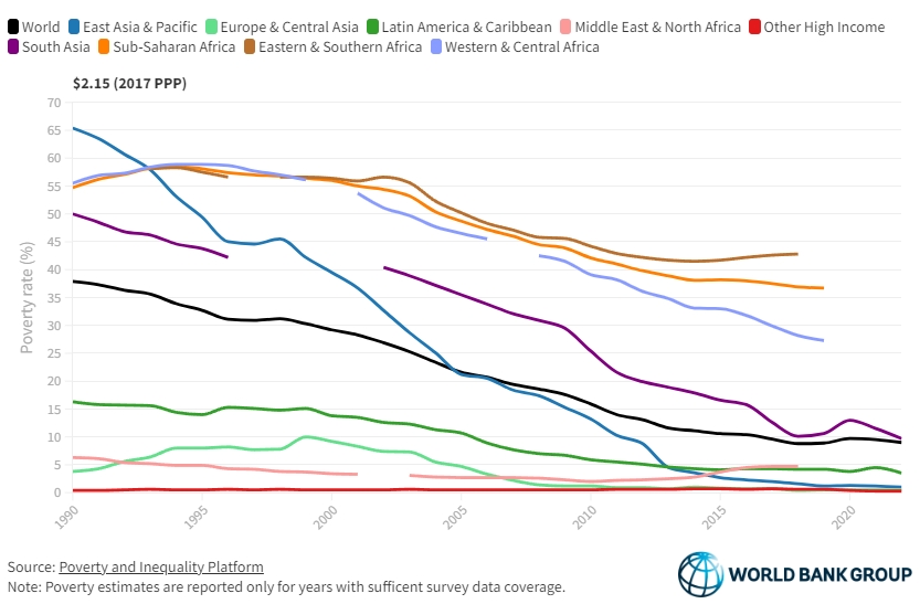
How did poverty rates decline so steeply? The reasons lie, to a large extent, in technological innovation, especially in genetics and farm management, and in the increased apport of plentiful and affordable meat protein to the world. The numbers below build an image of an industry that produces better, more, and cheaper.

Global meat production: 1950 vs. Present

Then…

In 1950, the estimated total meat production was of approximately 45 million metric tons.

Key Producers: The United States, Europe, and the Soviet Union were the primary producers of meat.
Types of Meat: Production was largely dominated by beef and pork, with poultry being less significant.

…and now

Now, the total meat production lies somewhere around 357 million metric tons (as of recent data from FAO)., representing a 53% increase from 2000 and a staggering 690% increase from 1950.

Key Producers: Major producers include China, the United States, Brazil, and the European Union.
Types of Meat: Significant increases in poultry production, with pork remaining a leading source of meat, especially in Asia. Beef production has also increased, but at a slower rate than poultry and pork.

Factors contributing to increased meat production

Population Growth: The world population has grown from approximately 2.5 billion in 1950 to over 8 billion today, driving increased demand for meat.

Economic Growth and Urbanization: Rising incomes and urbanization have led to shifts in economic power and dietary preferences, with more people consuming higher quantities of meat, especially in developing countries.

Technological Advancements: Improvements in animal breeding, feed efficiency, and production systems have increased the efficiency and output of meat production.

Intensification of Livestock Production: The shift from extensive to intensive livestock production systems has allowed for higher meat yields per animal.

Global Trade: Expansion of global trade in meat and meat products has facilitated the growth of production in countries with comparative advantages in livestock farming.

Livestock yield increase, 1950 to the present

The increase in livestock yield for cattle, pigs, and chickens between 1950 and the present has been significant due to advances in breeding, nutrition, management practices, and technology.

Beef

1950s

  • Average Carcass Weight: In the 1950s, the average carcass weight of beef cattle was about 200 to 250 kilograms (440 to 550 pounds).
  • Dressing Percentage: The dressing percentage (the proportion of live weight that becomes carcass) was typically around 50-55%.

Present Day

  • Average Carcass Weight: Today, the average carcass weight of beef cattle is approximately 300 to 400 kilograms (660 to 880 pounds).
  • Dressing Percentage: The dressing percentage has improved to about 60-65%.

Increase in Beef Cattle Yield

  • Increase in Carcass Weight: The average carcass weight has increased by about 100 to 150 kilograms (220 to 330 pounds) per animal.
  • Improved Dressing Percentage: The dressing percentage has increased by about 5-10 percentage points, meaning a greater proportion of the live weight is converted into meat.

Dairy

1950s

  • Average Milk Yield per Cow: Approximately 2,000 to 3,000 liters per year, depending on the region.

Present Day

  • Average Milk Yield per Cow: Approximately 8,000 to 10,000 liters per year globally, with some countries like the United States achieving even higher averages of 10,000 to 12,000 liters per year.

Increase in Milk Yield:: Milk yield per cow has increased about 4-5 times due to genetic selection, improved nutrition, technological advancements, and better herd management.
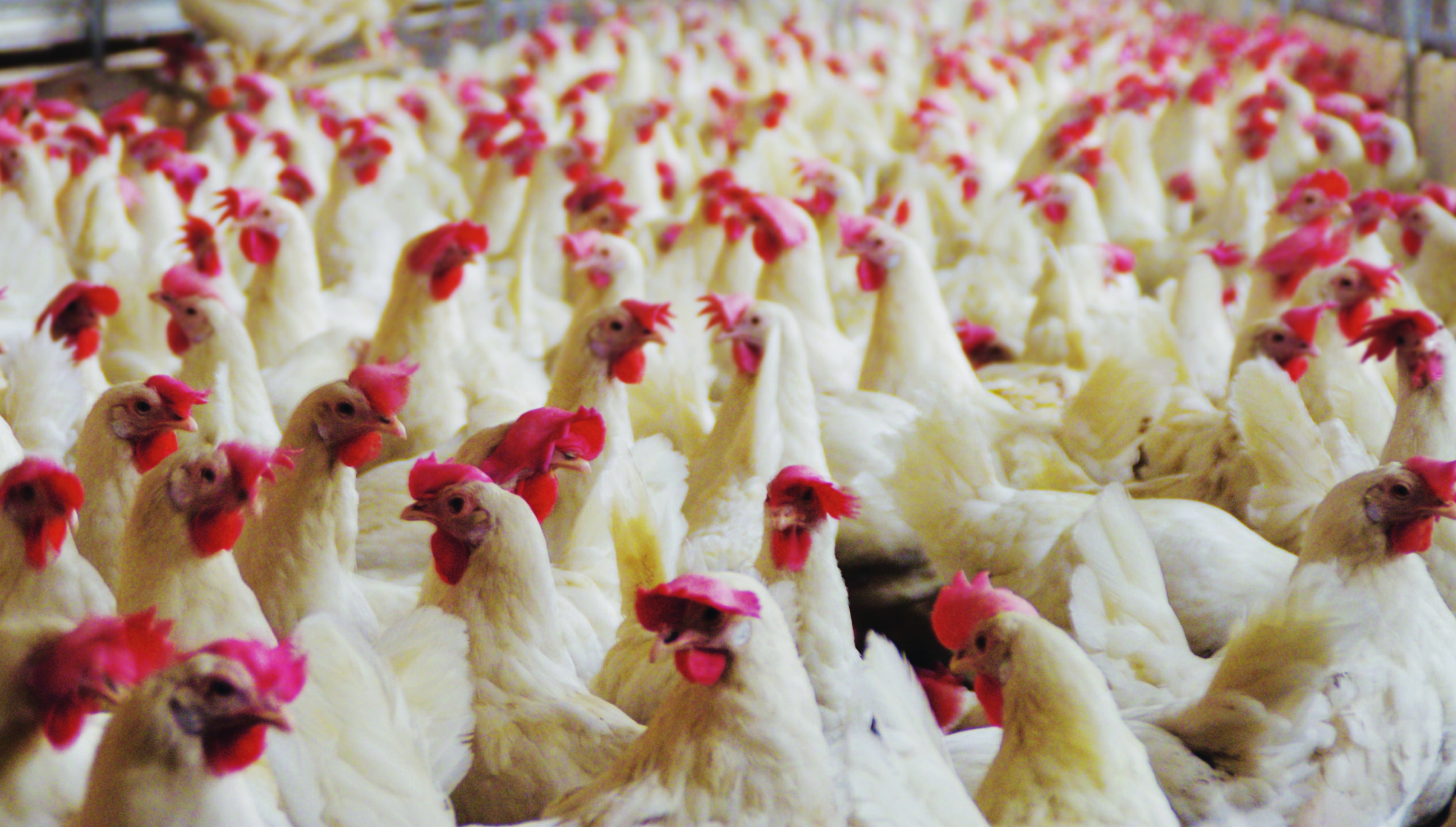
Chickens (Layers)

1950s

  • Average Egg Production per Hen: In the 1950s, a typical laying hen produced about 150 to 200 eggs per year.

Present Day

  • Average Egg Production per Hen: Today, a typical laying hen produces approximately 280 to 320 eggs per year, with some high-performing breeds producing even more.

Increase in Egg Yield: The average egg production per hen has increased by approximately 130 to 170 eggs per year.
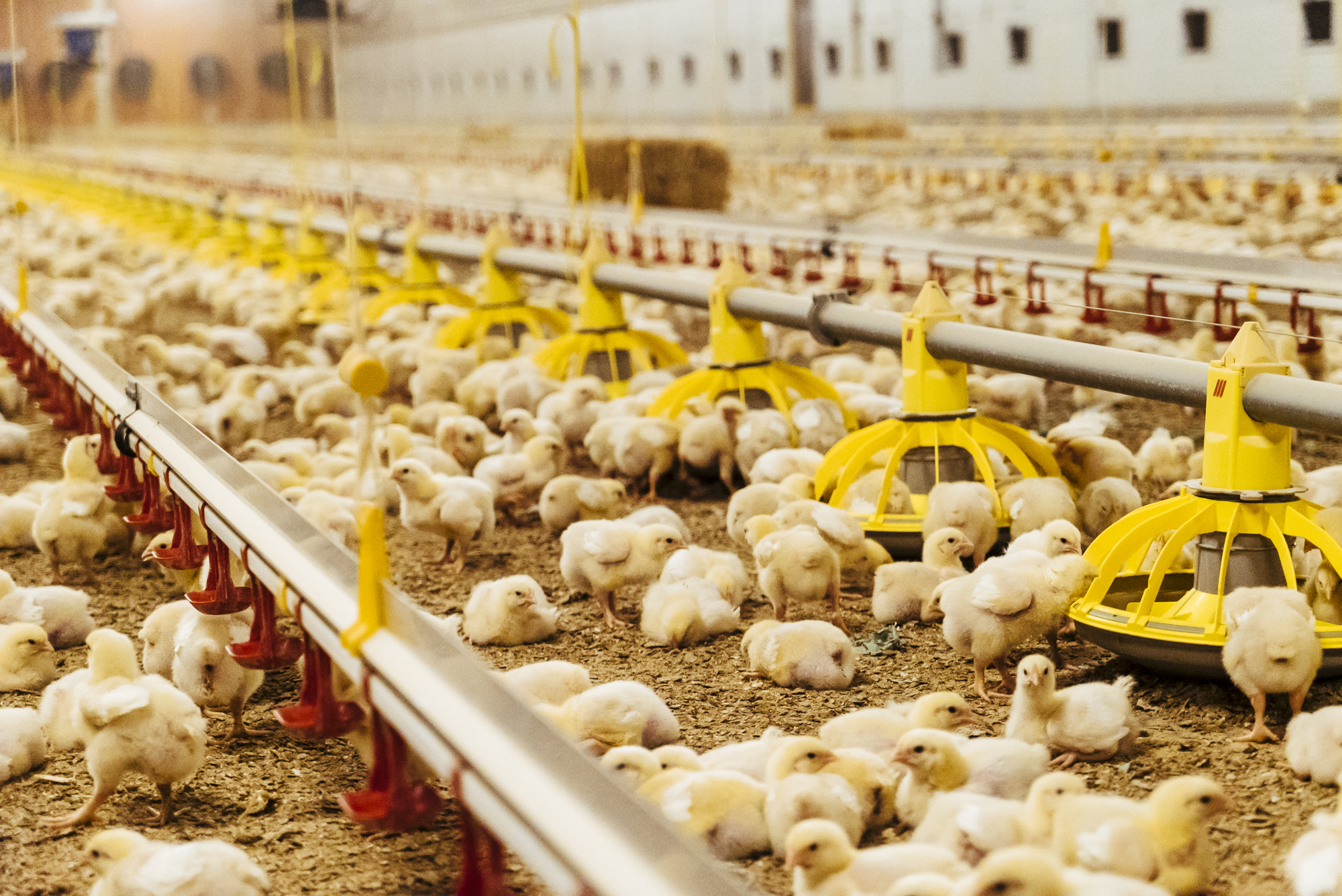
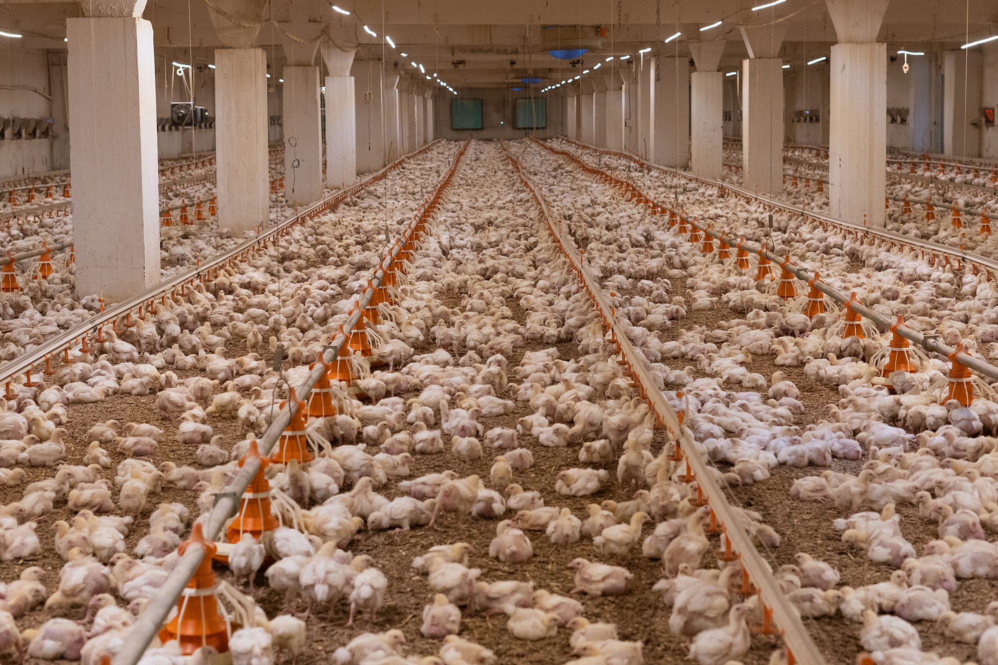
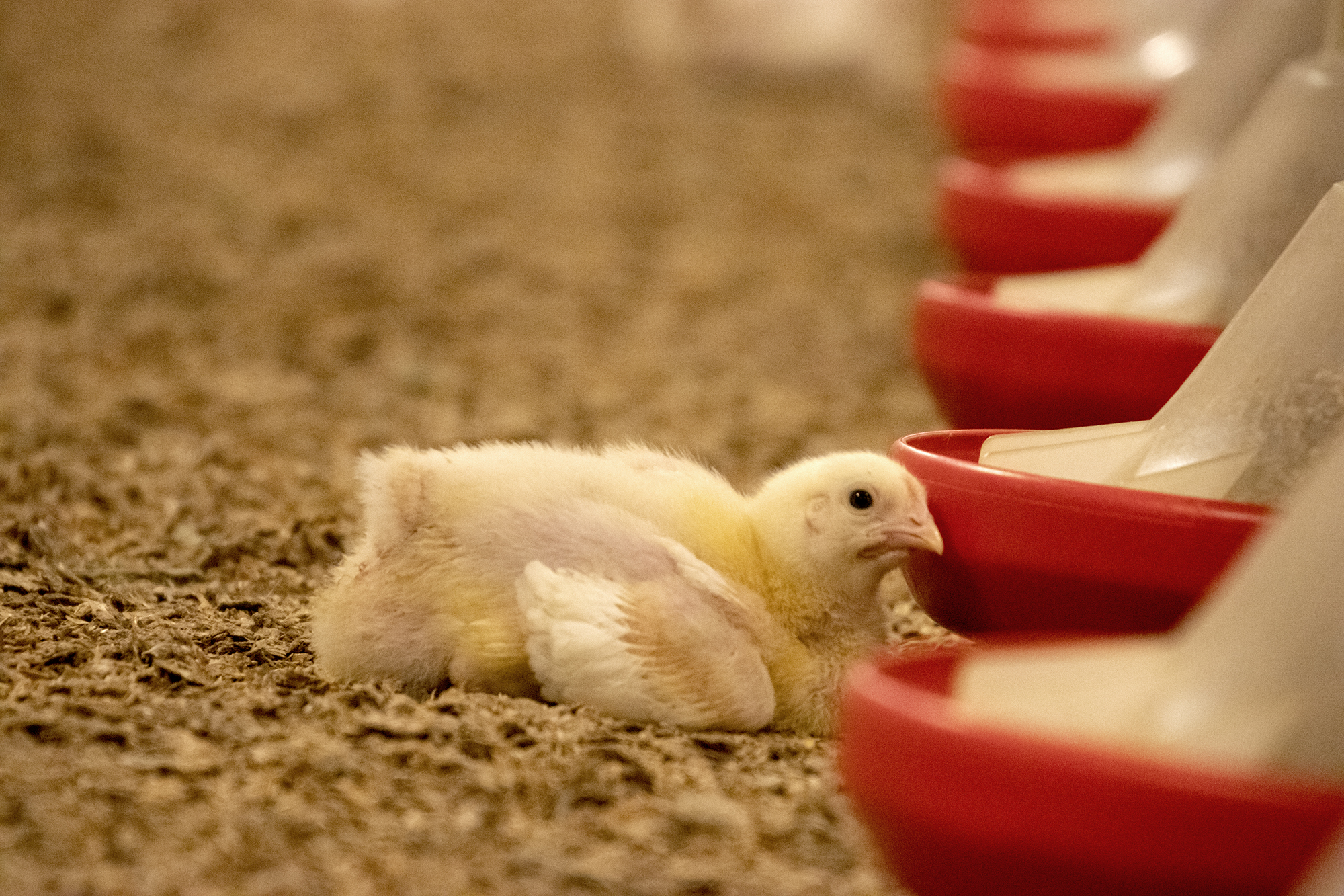
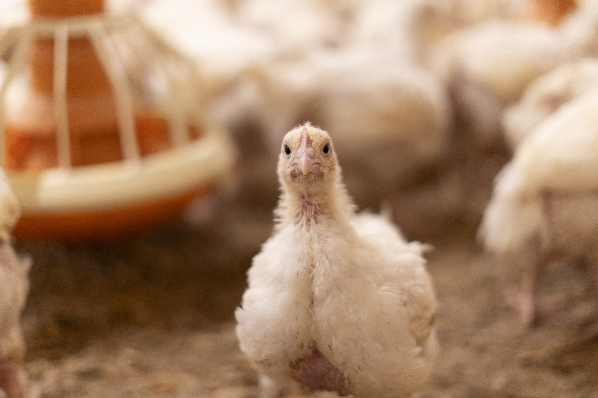
Chickens (Broilers)

1950s

  • Average Yield per Bird: In the 1950s, broiler chickens typically reached a market weight of about 1.5 to 2 kilograms (3.3 to 4.4 pounds) over a growth period of 10 to 12 weeks.

Present Day

  • Average Yield per Bird: Today, broiler chickens reach a market weight of about 2.5 to 3 kilograms (5.5 to 6.6 pounds) in just 5 to 7 weeks.

Increase in Yield: The average weight of a broiler chicken has increased by approximately 1 to 1.5 kilograms (2.2 to 3.3 pounds) per bird. Additionally, the time to reach market weight has been nearly halved.

Factors contributing to yield increases

Genetic Improvement:

  • Selective Breeding: Focused breeding programs have developed chicken strains with rapid growth rates and high feed efficiency, significantly increasing meat yield.

Nutrition:

  • Optimized Feed: Advances in poultry nutrition have led to feed formulations that promote faster growth and better health, using balanced diets rich in energy, protein, and essential nutrients.

Management Practices:

  • Housing and Environment: Improved housing conditions, including temperature and humidity control, have reduced stress and disease, enhancing growth rates.

Technological Advancements:

  • Automation: Automation in feeding, watering, and waste management has improved efficiency and bird health.
  • Health Monitoring: Advances in health monitoring and veterinary care have reduced mortality rates and supported faster growth.

Feed Conversion Efficiency:

  • Improved Feed Conversion Ratios (FCR): The amount of feed required to produce a unit of meat has decreased significantly, making production more efficient.

Why Feed Conversion Ratio is a sustainability metric

Feed Conversion Ratio (FCR) is a critical metric in livestock production that measures the efficiency with which animals convert feed into body mass. It is expressed as the amount of feed required to produce a unit of meat, milk, or eggs. Advances in nutrition and precision feeding allow producers to tailor diets that optimize FCR, reducing waste and improving nutrient uptake. Also, breeding programs focused on improving FCR can lead to livestock that naturally convert feed more efficiently, supporting long-term sustainability.

Poultry (Broilers): From the 1950s, improved from approximately 4.75 kg/kg to 1.7 kg/kg.

Pigs: From the 1950s, improved from about 4.5 kg/kg to 2.75 kg/kg.

Cattle (Beef): From the 1950s, improved from around 7.5 kg/kg to 6.0 kg/kg.

FCR ChangeFigure 4. Evolution of FCR from 1950

FCR is crucial for livestock sustainability for several reasons, as shown below.

1. Resource efficiency

Feed Costs: Feed is one of the largest operational costs in livestock production. A lower FCR means less feed is needed to produce the same amount of animal product, reducing costs and improving profitability.

Land Use: Efficient feed conversion reduces the demand for land needed to grow feed crops, helping to preserve natural ecosystems and decrease deforestation pressures.

Water Use: Producing less feed per unit of animal product reduces the water needed for crop irrigation, which is crucial in regions facing water scarcity.

2. Environmental impact

Greenhouse Gas Emissions: Livestock production is a significant source of greenhouse gases (GHGs), particularly methane from ruminants and nitrous oxide from manure management. Improved FCR means fewer animals are needed to meet production goals, reducing total emissions.

Nutrient Runoff: Efficient feed use minimizes excess nutrients that can lead to water pollution through runoff and eutrophication of aquatic ecosystems.

3. Animal welfare

Health and Growth: Optimizing FCR often involves improving animal health and growth rates, which can lead to better welfare outcomes. Healthy animals grow more efficiently and are less susceptible to disease.

4. Economic viability

Competitiveness: Lowering FCR improves the economic viability of livestock operations by reducing input costs and increasing competitiveness in the global market.

Food Security: Efficient livestock systems contribute to food security by maximizing the output of animal protein relative to the input of resources.

Improving FCR is essential for achieving sustainability in livestock production. It leads to more efficient resource use, reduced environmental impact, enhanced economic viability, and supports the well-being of animals. As global demand for animal products continues to rise, optimizing FCR will be crucial in balancing production with the need to protect and preserve natural resources.

“The eight warmest years on record since 1961 (and in fact since the beginning of observations in 1880) are all within the eight-year period of 2015–2022. Europe is the region where the temperature change has been the highest in 2022 (and also for most of the 2000–2022 period), with 2.23 °C, followed by Asia (1.75 °C), the Americas (1.05 °C), Africa (1.01 °C) and Oceania (0.8 °C). The average temperature change in the 2010s was 1.25 °C, compared to 0.96 °C in the 2000s.”
FAOSTAT 2023

Livestock emissions

Livestock emissions can be direct (farm-gate) or indirect (land use). Pre- and post-production emissions are considered separately, since they refer to emissions from manufacturing, processing, packaging, transport, retail, household consumption, and waste disposal.

GHG Emissions
Figure 5. Source: FAO

Farm-gate emissions

Global farm-gate emissions (related to the production of crops and livestock) grew by 14% between 2000 and 2021, to 7.8 Gt CO2 eq, see below. 53% come from livestock-related activities, and the emissions from enteric fermentation generated in the digestive system of ruminant livestock were alone responsible for 37 percent of agricultural emissions (FAOSTAT 2023).

World Farm Gate GHG Emissions By Activity
Figure 6. Source: FAO

Land use for livestock

Land use emissions contribute a large share to agricultural emissions overall, especially through deforestation (~74% of land-use GHG emissions). The numbers have declined in recent years, to a total of 21% reduction between 2000 and 2018.

The other side of the coin is represented by the increased land usage for livestock, either directly for grazing or indirectly for feed crops.

1. Pasture and grazing land

1950: Approximately 3.2 billion hectares (7.9 billion acres) were used as permanent pastures.

Present: The area has increased to around 3.5 billion hectares (8.6 billion acres).

Change: An increase of about 0.3 billion hectares (0.7 billion acres).

2. Land for Feed Crops

1950: The land area dedicated to growing feed crops (such as corn and soy) was significantly less than today due to lower livestock production intensities and smaller scale operations. Feed crops likely accounted for about 200-250 million hectares of the cropland, although figures are evidently difficult to estimate.

Present: Of the approx. 5 billion hectares of land globally used for agriculture, about 1.5 billion hectares are dedicated to cropland.

The increase in cropland hectares is a direct consequence of the intensification of demand for livestock production. To keep these numbers in check, it is essential that producers strive to use as little feed as possible for as much meat yield as possible – and this directly relates to a key metric of the feed additive industry: Feed Conversion Ratio, mentioned above.

The role of food loss in livestock sustainability

The Food and Agriculture Organization (FAO) of the United Nations defines food loss as the decrease in quantity or quality of food resulting from decisions and actions by food suppliers in the chain, excluding retail, food service providers, and consumers. Food loss specifically refers to food that gets spilled, spoiled, or lost before it reaches the consumer stage, primarily taking place during production, post-harvest, processing, and distribution stages.

Food loss is currently estimated to be relatively stable over the last decades, at around 13%.

Key aspects of food loss

  1. Stages of Food Loss:
    • Production: Losses that occur during agricultural production, including damage by pests or diseases and inefficiencies in harvesting techniques.
    • Post-Harvest Handling and Storage: Losses that happen due to inadequate storage facilities, poor handling practices, and lack of proper cooling or processing facilities.
    • Processing: Losses during the processing stage, which may include inefficient processing techniques, contamination, or mechanical damage.
    • Distribution: Losses that occur during transportation and distribution due to poor infrastructure, inadequate packaging, and logistical inefficiencies.
  2. Quality and Quantity:
    • Quality Loss: Refers to the reduction in the quality of food, affecting its nutritional value, taste, or safety, which may not necessarily reduce its quantity.
    • Quantity Loss: Refers to the actual reduction in the amount of food available for consumption due to physical losses.
  3. Exclusions:
    • Retail and Consumer Level: Food loss does not include food waste at the retail or consumer levels, which is categorized as food waste. Food waste refers to the discarding of food that is still fit for consumption by retailers or consumers.

Importance of reducing food loss

Every step along the production chain, each action taken to preserve feed, increase yield, ensure stable and high meat quality, can contribute to reducing food loss and ensuring that animal protein production stays sustainable and feeds the world more efficiently.

  • Food Security: Reducing food loss can help improve food availability and access, particularly in regions where food scarcity is a concern. Where we thought we were on our way to eradicate world hunger, recent upticks in several regions show us that progress is not a given.
  • Economic Efficiency: Minimizing food loss can improve the efficiency and profitability of food supply chains by maximizing the utilization of resources.
  • Environmental Impact: Reducing food loss helps to decrease the environmental footprint of food production by lowering greenhouse gas emissions and minimizing land and water use. This is all the more important in regions where world hunger shows signs of going up. Perhaps not by coincidence are these regions some of the most affected by climate change.

By understanding and addressing the causes of food loss, stakeholders across the food supply chain can work towards more sustainable and efficient food systems.

What’s next?

Improving production practices and technology

Investment in research and development of new technologies that enhance livestock production efficiency and reduce environmental impact is vital for the future sustainability of the sector.

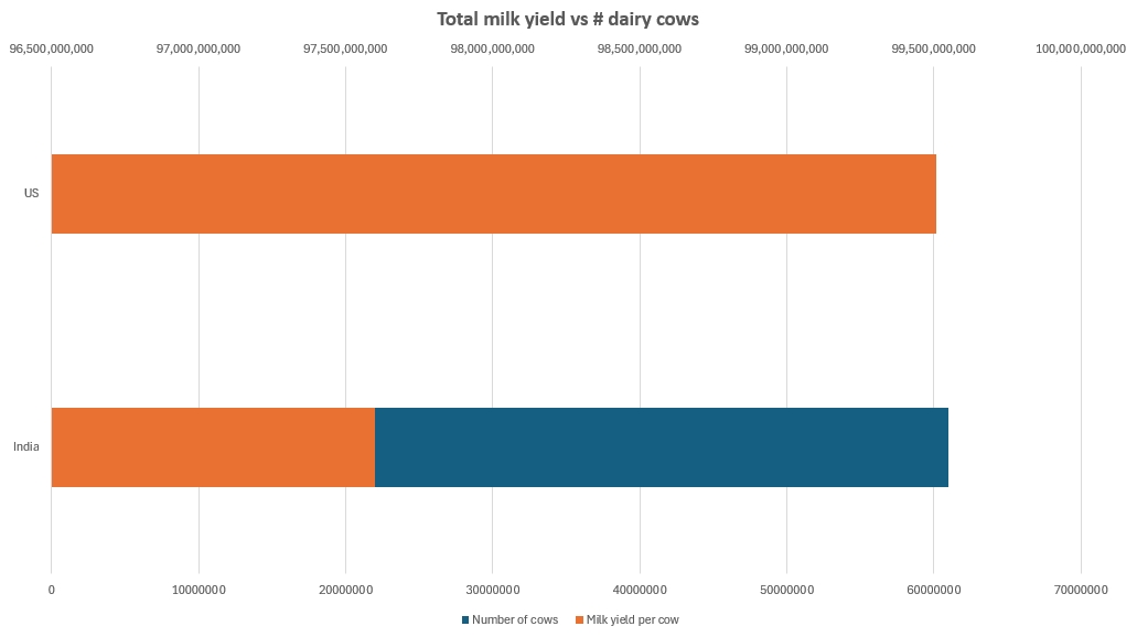
India is a good illustration of room to grow. If we look at cow milk alone, India, with a headcount of approximately 61 million animals, has a total milk production that is neck-and-neck with the United States, whose dairy cow headcount is in the neighborhood of 9.3 million. India’s milk yield sits around 1,600 liters/animal/year, compared to the US’s average of 10,700 liters.

Milk Yield IN US
Figure 7. Based on Our World in Data

Optimizing Feed Efficiency

Continued focus on improving FCR through genetic selection, optimized nutrition, and advanced management practices will be crucial for reducing the environmental footprint of livestock production.

Promoting Sustainable Land Use

Strategies to balance the need for increased livestock production with sustainable land use practices are essential. This includes adopting agroecological approaches and improving the efficiency of feed crop production.

Reducing Food Loss

Stakeholders across the food supply chain must prioritize reducing food loss through improved storage, transportation, and processing technologies. This will help ensure that livestock production contributes effectively to global food security.

Enhancing Emission Tracking and Reporting

There is a need for standardized methods for collecting and reporting data on GHG emissions in agriculture. This will enable more accurate assessments and the development of targeted strategies for emission reductions.

References

Bell, D. D. (2002). Laying hens in the U.S. market: An appraisal of trends from the beginning of the 20th century to present. Poultry Science, 81(5), 485-490. https://doi.org/10.1093/ps/81.5.485

CarbonWise. (2023). Global greenhouse gas emissions by sector. Retrieved from https://carbonwise.co/global-greenhouse-gas-emissions-by-sector/

Crippa, M., Solazzo, E., Guizzardi, D., Monforti-Ferrario, F., Tubiello, F. N., Leip, A., … & Janssens-Maenhout, G. (2022). Greenhouse gas emissions from food systems: building the global food system emissions database (GFED). Earth System Science Data, 14(4), 1795-1821. https://essd.copernicus.org/articles/14/1795/2022/essd-14-1795-2022.pdf

European Environment Agency (EEA). (2023). Improving the climate impact of raw material sourcing. Retrieved from https://www.eea.europa.eu/publications/improving-the-climate-impact-of-raw-material-sourcing

Food and Agriculture Organization of the United Nations (FAO). (2021). The State of Food and Agriculture 2021: Making agrifood systems more resilient to shocks and stresses. FAO. https://openknowledge.fao.org/server/api/core/bitstreams/6e04f2b4-82fc-4740-8cd5-9b66f5335239/content

Food and Agriculture Organization of the United Nations (FAO). (2021). Food Loss and Waste Database. FAO. https://www.fao.org/platform-food-loss-waste/food-loss/introduction/en

Food and Agriculture Organization of the United Nations (FAO). (2021). Greenhouse gas emissions from agrifood systems. Retrieved from https://www.fao.org/platform-food-loss-waste/food-loss/introduction/en

Goldewijk, K. K., & Verburg, P. H. (2013). Per-capita estimations of long-term historical land use and the consequences for global change research. Global Environmental Change, 23(4), 1166-1175. https://doi.org/10.1016/j.gloenvcha.2013.04.001

Intergovernmental Panel on Climate Change (IPCC). (2023). AR6 Synthesis Report: Climate Change 2023. IPCC. https://www.ipcc.ch/report/ar6/syr/

Kusuma, A. B., Laga, W. R., & Purnomo, H. (2022). Climate Change and Livestock Farming: Strategies for Mitigation and Adaptation. MDPI, 12(10), 1554. https://www.mdpi.com/2077-0472/12/10/1554

Matthews, D. (2023). Chicken, meat, and the future of global food: Forecasts and predictions for beef, pork, and more. Vox. https://www.vox.com/future-perfect/2023/8/4/23818952/chicken-meat-forecast-predictions-beef-pork-oecd-fao?mc_cid=d1a37e53b6&mc_eid=1b5c5e908a

Our World in Data. (2020). Milk yields per animal. Retrieved from https://ourworldindata.org/grapher/milk-yields-per-animal

Our World in Data. (2023). Grazing land use over the long-term, 1600 to 2023. Retrieved from https://ourworldindata.org/grazing-land-use-over-the-long-term

Ritchie, H., & Roser, M. (2020). Food greenhouse gas emissions. Our World in Data. https://ourworldindata.org/food-ghg-emissions

Roche, J. R., Friggens, N. C., Kay, J. K., Fisher, M. W., Stafford, K. J., & Berry, D. P. (2013). Invited review: Body condition score and its association with dairy cow productivity, health, and welfare. Animal Frontiers, 3(4), 23-29. https://doi.org/10.2527/af.2013-0032

Sharma, V. P., & Gulati, A. (2020). Changes in Herd Composition a Key to Indian Dairy Production. United States Department of Agriculture (USDA) Economic Research Service. https://www.ers.usda.gov/publications/pub-details/?pubid=99794

The Last Glaciers. (2023). Decarbonizing Food and Agriculture. Retrieved from https://thelastglaciers.com/decarbonising-food-and-agriculture/

Thoma, G., Jolliet, O., & Wang, Y. (2016). National Pork Board. (2016). Greenhouse gas emissions and the potential for mitigation from the pork industry in the U.S. Retrieved from https://www.porkcheckoff.org/wp-content/uploads/2021/05/16-214-THOMA-final-rpt.pdf

Thornton, P. K., & Herrero, M. (2015). Impacts of climate change on the livestock food supply chain; a review of the evidence. Frontiers in Veterinary Science, 2, 93. https://www.ncbi.nlm.nih.gov/pmc/articles/PMC4686767/

USDA – National Agricultural Statistics Service. (n.d.). Trends in U.S. Agriculture – Broiler Industry. U.S. Department of Agriculture. Retrieved from https://www.nass.usda.gov/Publications/Trends_in_U.S._Agriculture/Broiler_Industry/

Zuidhof, M. J., Schneider, B. L., Carney, V. L., Korver, D. R., & Robinson, F. E. (2014). Evolution of the modern broiler and feed efficiency. Annual Review of Animal Biosciences, 2(1), 47-71. https://doi.org/10.1146/annurev-animal-022513-114132


Global antimicrobial use in livestock farming: A revised estimate


Antimicrobial resistance (AMR) poses a significant threat to global health, driven by the overuse and misuse of antibiotics in both human medicine and livestock farming. In livestock farming, antimicrobials are still used extensively for therapeutic and non-therapeutic purposes. However, estimates of the quantities used per species are notoriously hard to derive from fragmented, incomplete, or unstandardized data around the world.

A recent article (“Global antimicrobial use in livestock farming: an estimate for cattle, chickens, and pigs”, Animal, 18(2), 2024) attempts to update the figures by estimating global biomass at treatment of cattle, pigs, and chickens, considering distinct weight categories for each species in biomass calculation, and using the European Medicines Agency’s weight standards for the animal categories. With these more refined calculations, authors Zahra Ardakani, Maurizio Aragrande, and Massino Canali aim to provide a more accurate estimate of global antimicrobial use (AMU) in cattle, chickens, and pigs. Understanding these patterns is crucial for addressing AMR and developing strategies for sustainable livestock management.

Key Findings

The study estimates that the global annual AMU for cattle, chickens, and pigs amounts to 76,060 tons of antimicrobial active ingredients. This is a significant revision from previous estimates due to a more detailed evaluation of animal weights and categories:

1. Cattle: 40,697 tons (53.5% of total AMU)
2. Pigs: 31,120 tons (40.9% of total AMU)
3. Chickens: 4,243 tons (5.6% of total AMU)

Figure 1: Distribution of global antimicrobial use among cattle, pigs, and chickens.

Methodology

The study utilizes the concept of Population Correction Units (PCU) to estimate antimicrobial usage, taking into account the weight and category of livestock at the time of treatment. This method differs from previous approaches that relied on live weight at slaughter, providing a more accurate representation of AMU.

The PCU is calculated by multiplying the number of animals by their average weight during treatment. This approach allows for differentiation by age and sex, which is particularly important for species like cattle and pigs.

Figure 2: (a) Changes in global PCU (million tonnes), (b) changes in global antibiotic use in mg per PCU, and (c) changes in global AMU (thousand tonnes) for cattle, chickens, and pigs; between 2010 and 2020.  Abbreviations: PCU = Population Correction Unit; AMU = Antibiotic Use.

Figure 2: (a) Changes in global PCU (million tonnes), (b) changes in global antibiotic use in mg per PCU, and (c) changes in global AMU (thousand tonnes) for cattle, chickens, and pigs; between 2010 and 2020. Abbreviations: PCU = Population Correction Unit; AMU = Antibiotic Use.

Study shows lower AMU than previous estimates

The study highlights a significant shift in AMU patterns, with chickens showing a remarkable decrease in antimicrobial use despite increased production. This is indicative of improved management and more responsible use of antibiotics in the poultry industry.

The lower AMU in cattle and pigs, compared to previous estimates, underscores the importance of considering animal age and weight at treatment. These findings align closely with World Organization for Animal Health (WOAH) estimates, validating the methodology.

However, the study also acknowledges limitations, including reliance on European standards for average weight at treatment, which may not reflect global variations. Additionally, the lack of comprehensive global data on veterinary antibiotics presents challenges in creating fully accurate estimates.

Corrected estimate highlights improved production advances

This study provides a revised and potentially more accurate estimate of global antimicrobial use in livestock. By accounting for the weight and treatment categories of animals, it offers insights that could guide policy and management practices to mitigate the spread of antimicrobial resistance.

The article also indicates that the industry may have over-estimated antimicrobial usage in livestock and, just as importantly, that antimicrobial use has been kept in check or even reduced, despite increases in farmed animal headcounts. The lower usage is likely due to regulatory oversight and improvements in alternative methods to control and mitigate health challenges.


Mycotoxins in poultry – External signs can give a hint

Part 4: Paleness

By Dr. Inge Heinzl, Editor and Marisabel Caballero, Global Technical Manager Poultry

We already showed bad feathering, mouth and beak lesions, bone issues, and foot pad lesions as signs of mycotoxin contamination in the feed, but there is another indicator: paleness. Paleness can signify a low count of red blood cells resulting from blood loss or inadequate production of these cells. Other possibilities are higher bilirubin levels in the blood due to an impaired liver, leading to jaundice or missing pigmentation.

Hen With Pale Comb And Wattles Large
Hen with pale comb and wattles (adapted from Bozzo et al., 2023)

The mycotoxins mainly causing anemia are Aflatoxins, Ochratoxin, DON, and T-2 toxin

Anemia can be diagnosed using parameters such as red blood cell count, hemoglobin levels, and hematocrit/packed cell volume (PCV). Numerous studies have examined the impact of mycotoxins on hematological parameters. They reveal their propensity to affect red blood cell production by impairing the function of the spleen and inducing hematological alterations. On the other hand, anemia can be caused by blood loss. Due to affecting coagulation factors, mycotoxins can lead to internal hemorrhages. The gut wall damage, probably due to secondary infections such as coccidiosis and necrotic enteritis, can entail bloody diarrhea in various animal species.

Impact on the production of blood cells

Low values of blood parameters such as red blood cells, hemoglobin, and hematocrit can result from inadequate production due to impacted production organs. The World Health Organization (WHO, 1990) and European Commission (European Commission, 2001) have identified hematopoietic tissues as targets for necrosis caused by T-2 toxin. Chu (2003) even stated that “the major lesion of T-2 toxin is its devastating effect on the hematopoietic system in many mammals, including humans”. Pande et al. (2006) suggested that reduced hemoglobin values result from decreased protein synthesis due to mycotoxin contamination, a notion supported by Pronk et al. (2002), who described trichothecenes as potent inhibitors of protein, DNA, and RNA synthesis, particularly affecting tissues with high cell division rates. Additionally, the European Commission (2001) highlighted the sensitivity of red blood cell progenitor cells (in this trial, the cells of mice, rats, and humans) to the toxic effects of T-2 and HT-toxins. DAS also seems to attack the hematopoietic system, as shown in humans (WHO, 1990). A further cause for anemia might be low feed intake or nutrient absorption, which inhibits adequate iron absorption and leads to iron deficiency. In their case report, Bozzo et al. (2023) assumed that renal failure and a resulting impaired excretion capacity caused by OTA might even increase the half-life of the toxins. This would enhance their effects on their target organs, such as the liver and bone marrow, and lead to anemia.

Several studies utilizing different animal species and mycotoxin dosages have been conducted to assess the effects of Aflatoxins, Ochratoxin, and T-2 Toxin on hematological parameters. The following table provides a summary of some of these studies.

Animal species Dosage Impact Reference
T-2 Toxin and other Trichothecenes
Broilers T-2 – 0, 1, 2, and 4 mg T-2 toxin/kg

n=30 per group

Significant reduction in hemoglobin at 1, 2, and 4 ppm; PCV significantly reduced at 4 ppm Pande et al., 2006
Broilers T-2 – 0 and 4 mg/kg diet

n=60 per group

Decrease in hemoglobin, mean corpuscular volume, and mean corpuscular hemoglobin concentration Kubena et al., 1989a
Broilers 4, 16, 50, 100, 300 ppm for seven days

n=5-20 chickens per group

Anemia; significant reduction of hematocrit (50 and 100 ppm); survivors had atrophied lymphoid organs and were anemic Hoerr et al., 1982
Yangzhou goslings 0, 0.2, 0.4, 0.6, 0.8, 1.0, 2.0 mg/kg; n=6 per group Red blood cell count decreased in the 2.0 mg/kg group along with an increase in mean corpuscular hemoglobin (p<0.05) and reduced mean platelet volume (P<0.05) Gu et al., 2023
Broilers 2 ppm; 32 birds per group Anemia, as indicated by significantly (P<0.05) lower total erythrocyte count (TEC) values, lower hemoglobin levels, and packed cell volume; additional thrombocytopenia could be the cause of bleeding Yohannes et al., 2013
DON
Broilers 5 and 15 mg/kg of feed for 42 days Decrease in erythrocytes, mean corpuscular volume (MCV), and mean corpuscular hemoglobin concentration (MCHC) at 15 mg/kg; decrease in hematocrit and hemoglobin at both levels of DON.


Riahi, 2021
Piglets 0.6 mg/kg and 2.0 mg/kg Significant decrease in mean corpuscular volume Modrá et al., 2013
Broilers 16 mg/kg diet

n=60 per group

Significant decrease in mean corpuscular volume Kubena et al., 1989c
Ochratoxin
Broilers 2 mg/kg diet singly or combined with

DAS 6 mg/kg

Reduced mean corpuscular hemoglobin values Kubena et al., 1994
Broilers 2 mg/kg diet Significant decrease in hemoglobin, hematocrit, mean corpuscular volume and mean corpuscular hemoglobin concentration Kubena et al., 1989b
Aflatoxins
Broilers 2.5 µg/g Decrease in red blood cell count Huff et al., 1988
Broilers ≥1.25 µg/g Significant decrease in hemoglobin and erythrocyte count Tung et al., 1975
AFB1 + OTA
Laying hens Natural feed contamination OTA – 31 ± 3.08 µg/kg and

AFB1 – 5.6 ± 0.33 µg/kg dry weight

Anemia signs (pale appearance of combs and wattles), evidenced by the discoloration of the content of the femoral medullary cavity.


Bozzo et al., 2023


Table 1: The effects of different mycotoxins on hematological parameters – hematopoiesis

In their meta-analysis, Andretta et al. (2012) reported that the presence of mycotoxins in broiler diets decreased the hematocrit and the hemoglobin concentration by 5% and 15%, and aflatoxin alone decreased the parameters by 6% and 20%.

It should be evident that a simultaneous occurrence of several mycotoxins even aggravates the situation. In an experiment involving Sprague Dawley rats, administering T-2, DON, NIV, ZEA, NEO, and OTB decreased hematocrit and red blood cell counts across all mycotoxins. However, for DON, NIV, ZEN, and OTB, red blood cell values showed partial recovery after 24 hours (Chattopadhyay, 2013). Perhaps the organism learns to cope with the mycotoxins.

The examples show that Trichothecenes, such as T-2 toxin, DON, and others, as well as Ochratoxins and Aflatoxins, impact blood parameters such as hematocrit, hemoglobin, red blood cell count, and mean corpuscular volume. All these changes might lead to paleness of the skin and birds’ feet and combs.

Blood loss caused by bleeding or destruction of erythrocytes

The second possibility for anemia is blood loss due to injuries or lesions. In addition to directly causing hemorrhages, mycotoxins can promote secondary infections such as coccidiosis, which damages the gut and may produce bloody feces.

Parent-Massin (2004) e.g. reports on rapidly progressing coagulation problems after the ingestion of trichothecenes leading to septicemia and massive hemorrhages. Table 2 shows more examples of mycotoxins causing paleness due to blood loss.

Animal species Dosage Impact Reference
T-2 Toxin and other Trichothecenes
Cats T-2 toxin – 0.06-0.1 mg/kg body weight/day Bloody feces, hemorrhages Lutsky et al., 1978
Cats T-2 toxin – 0.08 mg/kg BW every 48 h until death Bloody feces Lutzky and Mor, 1981
Pigeon DAS in oat, sifting Emesis and bloody stools Szathmary (1983)
Calves 0.08, 0.16, 0.32, or 0.6 mg/kg BW per day for 30 days; 1 calf per treatment Bloody feces at doses ≥0.32 mg/kg BW per day Pier et al., 1976
Ochratoxin
Rats Single dosages of 0, 17, or 22 mg/kg BW in 0.1 Mol/L NaHCO3, gavage Multifocal hemorrhages in many organs Albassam et al., 1987
 
DON
Broilers 0, 35, 70, 140, 280, 560, and 1120 mg/kg body weight Ecchymotic hemorrhages throughout the intestinal tract, liver, and musculature; relationship to hemorrhagic anemia syndrome seems warranted Huff et al., 1981
Sterigmatocystin (ST)
10-12-day old chicks (93-101 g) 10 and 14 mg/kg BW intraperitoneal Hemorrhages and foci of necrosis in the liver Sreemannarayana et al., 1987
Aflatoxins
Broiler chickens 100 µg/kg feed Hemorrhages in the liver Abdel-Sattar, 2019
Turkeys 500 and 1000 ppb in the diet Bloody diarrhea, spleens with hemorrhages, petechial hemorrhages in the small intestine Giambrone et al., 1984
Broilers 0, 0.625, 1.25, 2.5, 5.0, and 10.0 mg/kg of diet combined with Infectious Bursal Disease Slight hemorrhages in the skeletal muscles; decreased hematocrit and hemoglobin due to hemolytic anemia. Chang and Hamilton, 1981
Broilers 0, 1, and 2 mg AFB1/kg of diet Downregulation of the genes involved in blood coagulation (coagulation factor IX and X) and upregulation of anticoagulant protein C precursor, an inactivator of coagulation factors Va and VIIIa, and antithrombin-III precursor with 2 mg/kg Yarru, 2009
Pigs 1-4 mg/kg, 4 weeks

0.4-0.8 mg/kg, 10 weeks

Hemorrhages Henry et al., 2001

Table 2: The effects of different mycotoxins on hematological parameters – blood loss

Poor pigmentation

The fourth reason for paleness can be inadequate pigmentation. According to Hy Line (2021), the so-called pale bird syndrome is characterized by poor skin and egg yolk pigmentation and is caused by reduced absorption of fat and carotenoid pigments in compromised birds. This is also the case when the diets contain pigment supplements. Tyczkowski and Hamilton (1986) observed in their experiment with chickens exposed to doses of 1-8 µg of Aflatoxins/g of diet for three weeks that aflatoxins can cause poor pigmentation in chickens, probably by impairing carotenoids absorption but also transport and deposition. Osborne et al. (1982) asserted that carotenoids were significantly (P<0.05) depressed by 2 ppm ochratoxin as well as by 2.5 ppm aflatoxin in the diet.

Another possibility is oxidative stress due to the mycotoxin challenge. As pigments also serve as antioxidants, they may be expended for this purpose and are no longer available for pigmentation.

Paleness in poultry – a reason to think about mycotoxins

Paleness can have different causes, some of which are influenced by mycotoxins. If your chickens or hens are pale, checking the feed concerning mycotoxins is always recommended. A feed analysis can give information about possible contamination (see our tool MasterRisk).

In the case of contamination, effective products binding the mycotoxins and mitigating the adverse effects of these harmful substances can help protect your birds. As paleness is usually not the only effect of mycotoxins but also a decrease in growth, toxin binders can help maintain the performance of your animals.

References:

Abdel-Sattar, Ward Masoud, Kadry Mohamed Sadek, Ahmed Ragab Elbestawy, and Disouky Mohamed Mourad. “The Protective Role of Date Palm (Phoenix Dactylifera Seeds) against Aflatoxicosis in Broiler Chickens Regarding Carcass Characterstics, Hepatic and Renal Biochemical Function Tests and Histopathology.” Journal of World’s Poultry Research 9, no. 2 (June 25, 2019): 59–69. https://doi.org/10.36380/scil.2019.wvj9.

Albassam, M. A., S. I. Yong, R. Bhatnagar, A. K. Sharma, and M. G. Prior. “Histopathologic and Electron Microscopic Studies on the Acute Toxicity of Ochratoxin a in Rats.” Veterinary Pathology 24, no. 5 (September 1987): 427–35. https://doi.org/10.1177/030098588702400510.

Andretta, I., M. Kipper, C.R. Lehnen, and P.A. Lovatto. “Meta-Analysis of the Relationship of Mycotoxins with Biochemical and Hematological Parameters in Broilers.” Poultry Science 91, no. 2 (February 2012): 376–82. https://doi.org/10.3382/ps.2011-01813.

Bhat, RameshV, Y Ramakrishna, SashidharR Beedu, and K.L Munshi. “Outbreak of Trichothecene Mycotoxicosis Associated with Consumption of Mould-Damaged Wheat Products in Kashmir Valley, India.” The Lancet 333, no. 8628 (January 1989): 35–37. https://doi.org/10.1016/s0140-6736(89)91684-x.

Bozzo, Giancarlo, Nicola Pugliese, Rossella Samarelli, Antonella Schiavone, Michela Maria Dimuccio, Elena Circella, Elisabetta Bonerba, Edmondo Ceci, and Antonio Camarda. “Ochratoxin A and Aflatoxin B1 Detection in Laying Hens for Omega 3-Enriched Eggs Production.” Agriculture 13, no. 1 (January 5, 2023): 138. https://doi.org/10.3390/agriculture13010138.

Chang, Chao-Fu, and Pat B. Hamilton. “Increased Severity and New Symptoms of Infectious Bursal Disease during Aflatoxicosis in Broiler Chickens.” Poultry Science 61, no. 6 (June 1982): 1061–68. https://doi.org/10.3382/ps.0611061.

Chattopadhyay, Pronobesh, Amit Agnihotri, Danswerang Ghoyary, Aadesh Upadhyay, Sanjeev Karmakar, and Vijay Veer. “Comparative Hematoxicity of Fusarium Mycotoxin in Experimental Sprague-Dawley Rats.” Toxicology International 20, no. 1 (2013): 25. https://doi.org/10.4103/0971-6580.111552.

European Commission. “Opinion of the Scientific Committee on Food on Fusarium Toxins Part 5: T-2 Toxin and HT-2 Toxin.” Food.ec.europa. Accessed May 30, 2001. https://food.ec.europa.eu/document/download/a859c348-a38e-404c-a2af-c3e29a3a8777_en?filename=sci-com_scf_out88_en.pdf.

Giambrone, J.J., U.L. Diener, N.D. Davis, V.S. Panangala, and F.J. Hoerr. “Effect of Purified Aflatoxin on Turkeys.” Poultry Science 64, no. 5 (May 1985): 859–65. https://doi.org/10.3382/ps.0640859.

Gu, Wang, Qiang Bao, Kaiqi Weng, Jinlu Liu, Shuwen Luo, Jianzhou Chen, Zheng Li, et al. “Effects of T-2 Toxin on Growth Performance, Feather Quality, Tibia Development and Blood Parameters in Yangzhou Goslings.” Poultry Science 102, no. 2 (February 2023): 102382. https://doi.org/10.1016/j.psj.2022.102382.

Henry, H., T. Whitaker, I. Rabban, J. Bowers, D. Park, W. Price, F.X. Bosch, et al. “Aflatoxin M1.” Aflatoxin M1 (JECFA 47, 2001). Accessed July 29, 2024. https://inchem.org/documents/jecfa/jecmono/v47je02.htm.

Hoerr, F., W. Carlton, B. Yagen, and A. Joffe. “Mycotoxicosis Caused by Either T-2 Toxin or Diacetoxyscirpenol in the Diet of Broiler Chickens.” Fundamental and Applied Toxicology 2, no. 3 (May 1982): 121–24. https://doi.org/10.1016/s0272-0590(82)80092-4.

Huff, W.E., J.A. Doerr, P.B. Hamilton, and R.F. Vesonder. “Acute Toxicity of Vomitoxin (Deoxynivalenol) in Broiler Chickens,” Poultry Science 60, no. 7 (July 1981): 1412–14. https://doi.org/10.3382/ps.0601412.

Huff, W.E., R.B. Harvey, L.F. Kubena, and G.E. Rottinghaus. “Toxic Synergism between Aflatoxin and T-2 Toxin in Broiler Chickens.” Poultry Science 67, no. 10 (October 1988): 1418–23. https://doi.org/10.3382/ps.0671418.

Hy-Line. “Mycotoxins: How to deal with the threat of mycotoxicosis.” Hy-Line International. Accessed July 29, 2024. https://www.hyline.com/.

Klein, P. J., T. R. Vleet, J. O. Hall, and R. A. Coulombe. “Dietary Butylated Hydroxytoluene Protects against Aflatoxicosis in Turkey.” Poisonous plants and related toxins, November 24, 2003, 478–83. https://doi.org/10.1079/9780851996141.0478.

Kubena, L.F., R.B. Harvey, T.S. Edrington, and G.E. Rottinghaus. “Influence of Ochratoxin A and Diacetoxyscirpenol Singly and in Combination on Broiler Chickens.” Poultry Science 73, no. 3 (March 1994): 408–15. https://doi.org/10.3382/ps.0730408.

Kubena, L.F., R.B. Harvey, W.E. Huff, D.E. Corrier, T.D. Philipps, and G.E. Rottinghaus. “Influence of Ochratoxin A and T-2 Toxin Singly and in Combination on Broiler Chickens.” Poultry Science 68, no. 7 (July 1989): 867–72. https://doi.org/10.3382/ps.0680867.

Kubena, L.F., R.B. Harvey, W.E. Huff, D.E. Corrier, T.D. Phillips, and G.E. Rottinghaus. “Influence of Ochratoxin A and T-2 Toxin Singly and in Combination on Broiler Chickens.” Poultry Science 68, no. 7 (July 1989): 867–72. https://doi.org/10.3382/ps.0680867.

Kubena, L.F., W.E. Huff, R.B. Harvey, T.D. Phillips, and G.E. Rottinghaus. “Individual and Combined Toxicity of Deoxynivalenol and T-2 Toxin in Broiler Chicks.” Poultry Science 68, no. 5 (May 1989): 622–26. https://doi.org/10.3382/ps.0680622.

Lutsky, I.I., and N. Mor. “Alimentary Toxic Aleukia (Septic Angina, Endemic Panmyelotoxicosis, Alimentary Hemorrhagic Aleukia): T-2 Toxin-Induced Intoxication of Cats.” The American journal of pathology, 1980. https://pubmed.ncbi.nlm.nih.gov/6973281/.

Lutsky, Irving, Natan Mor, Boris Yagen, and Avraham Z. Joffe. “The Role of T-2 Toxin in Experimental Alimentary Toxic Aleukia: A Toxicity Study in Cats.” Toxicology and Applied Pharmacology 43, no. 1 (January 1978): 111–24. https://doi.org/10.1016/s0041-008x(78)80036-2.

MEJ, Pronk, Schothorst RC, and H.P. van Egmond. “Toxicology and Occurrence of Nivalenol, Fusarenon X, Diacetoxyscirpenol, Neosolaniol and 3- and 15- Acetyldeoxynivalenol; a Review of Six Trichothecenes.” Home – Web-based Archive of RIVM Publications, November 7, 2002. https://rivm.openrepository.com/handle/10029/9184.

Modra, Helena, Jana Blahova, Petr Marsalek, Tomas Banoch, Petr Fictum, and Martin Svoboda. “The Effects of Mycotoxin Deoxynivalenol (DON) on Haematological and Biochemical Parameters and Selected Parameters of Oxidative Stress in Piglets.” Neuro Endocrinol Lett. 34, no. Suppl 2 (2013): 84–89.

Osborne, D.J., W.E. Huff, P.B. Hamilton, and H.R. Burmeister. “Comparison of Ochratoxin, Aflatoxin, and T-2 Toxin for Their Effects on Selected Parameters Related to Digestion and Evidence for Specific Metabolism of Carotenoids in Chickens,” Poultry Science 61, no. 8 (August 1982): 1646–52. https://doi.org/10.3382/ps.0611646.

Pande, Vivek, Nitin Kurkure, and A.G. Bhandarkar. “Effect of T-2 Toxin on Growth, Performance and Haematobiochemical Alterations in Broilers .” Indian Journal of Experimental Biology 44, no. 1 (February 2006): 86–88.

Pier , A.C., S.J. Cysewski, J.L. Richard , A.L. Baetz, and L. Mitchell. “Experimental Mycotoxicoses in Calves with Aflatoxin, Ochratoxin, Rubratoxin, and T-2 Toxin.” Proceedings, annual meeting of the United States Animal Health Association, 1976. https://pubmed.ncbi.nlm.nih.gov/1078072/.

Resanovic, Radmila, Ksenija Nesic, Vladimir Nesic, Todor Palic, and Vesna Jacevic. “Mycotoxins in Poultry Production.” Zbornik Matice srpske za prirodne nauke, no. 116 (2009): 7–14. https://doi.org/10.2298/zmspn0916007r.

Riahi, Insaf, Virginie Marquis, Anna Maria Pérez-Vendrell, Joaquim Brufau, Enric Esteve-Garcia, and Antonio J. Ramos. “Effects of Deoxynivalenol-Contaminated Diets on Metabolic and Immunological Parameters in Broiler Chickens.” Animals 11, no. 1 (January 11, 2021): 147. https://doi.org/10.3390/ani11010147.

Sreemannarayana, O., A. A. Frohlich, and R. R. Marquardt. “Acute Toxicity of Sterigmatocystin to Chicks.” Mycopathologia 97, no. 1 (January 1987): 51–59. https://doi.org/10.1007/bf00437331.

Stack, Jim, and Mike Carlson. “Fumonisins in Corn.” DigitalCommons@University of Nebraska – Lincoln, 2003. https://core.ac.uk/download/pdf/188054556.pdf.

Szathmary, C.I. “Trichothecene Toxicoses and Natural Occurrence in Hungary.” Essay. In Ueno, Y: Developments in Food Science IV. Trichothecenes, 229–50. New York: Elsevier, 1983.

Tung, Hsi-Tang, F.W. Cook, R.D. Wyatt, and P.B. Hamilton. “The Anemia Caused by Aflatoxin.” Poultry Science 54, no. 6 (November 1975): 1962–69. https://doi.org/10.3382/ps.0541962.

Tyczkowski, Juliusz K., and Pat B. Hamilton. “Altered Metabolism of Carotenoids during Aflatoxicosis in Young Chickens,” Poultry Science 66, no. 7 (July 1987): 1184–88. https://doi.org/10.3382/ps.0661184.

WHO. “Selected Mycotoxins : Ochratoxins, Trichothecenes, Ergot / Published under the Joint Sponsorship of the United Nations Environment Programme, the International Labour Organisation and the World Health Organization.” World Health Organization, January 1, 1990. https://apps.who.int/iris/handle/10665/39552.

Yohannes, T., A. K. Sharma, S. D. Singh, and V. Sumi. “Experimental Haematobiochemical Alterations in Broiler Chickens Fed with T-2 Toxin and Co-Infected with IBV.” Open Journal of Veterinary Medicine 03, no. 05 (2013): 252–58. https://doi.org/10.4236/ojvm.2013.35040.


Mycotoxins in poultry – External signs can give a hint

Part 3: Bone disorders and foot pad lesions

By Dr. Inge Heinzl, Editor, and Marisabel Caballero, Global Technical Manager Poultry


Bone health is essential for animals and humans. Besides giving structural support, allowing movement, and protecting vital organs, the bones release hormones that are crucial for mineral homeostasis and acid balance and serve as reservoirs of energy and minerals (Guntur & Rosen, 2012; Rath, N.C. & Durairaj, 2022; Suchacki et al., 2017).

Bone disorders and foot pad lesions are considerable challenges in poultry production, especially for fast-growing birds with high final weights. Due to pain, the animals do not move, and dominant, healthy birds may restrict lame birds’ access to feed and water. In consequence, these birds are often culled. Moreover, processing these birds is problematic, and often, they must be discarded or downgraded.

Foot pad lesions, another common issue in poultry production, can also have significant economic implications. On the one hand, pain restricts birds from eating and drinking and reduces weight gain. On the other hand, for many producers, chicken feet constitute a substantial part of the economic value of the bird; therefore, discarding them represents a significant financial loss. Additionally, to push poultry production in the right direction concerning animal health and welfare, a foot pad scoring system at the processing plant is in place in European countries.

Mycotoxins affect bones in different ways

Mycotoxins, depending on their target organs, can have diverse effects on the skeleton of birds. For example, mycotoxins that target the liver can disrupt calcium metabolism, which in turn affects the mineralization of the bones (rickets) and the impairment of chondrocytes can slow down bone growth (e.g., tibial dyschondroplasia). When the kidneys are impacted, urate clearance decreases, plasma uric acid consequently increases, and urate crystals form in the synovial fluid and tendon sheaths of various joints, particularly the hock joints. These examples highlight the complex and varied ways mycotoxins can impact poultry bone health.

Inadequate bone mineralization and strength – Rickets and layer cage fatigue

Sufficient bone mineralization is essential for the stability of the skeleton. Calcium (Ca), Vitamin D, and Phosphorous (P) deficiency leads to inadequate mineralization, weakens the bone, and can cause soft and bent bones or, in the case of layers, cage fatigue – a collapse of the spinal bone- and paralysis. Inadequate bone mineralization can be caused in different ways, among them:

  1. Decrease in the availability of the nutrients necessary for mineralization. This can occur if the digestibility of these nutrients deteriorates
  2. Impact on the Ca/P ratio—A ratio of 1 – 2:1 is vital for adequate bone development (Loughrill et al., 2016). Mycotoxins can alter absorption and transporters for one or both elements, altering their ratio.
  3. Impact on the Vitamin D receptor, affecting its expression or the transporters for Ca and P.

Aflatoxins can impair bone mineralization by different modes of action. An important one is the impairment of the digestibility of Ca and P: Kermanshahi et al. (2007) fed broilers diets with high levels of aflatoxins (0.8 to 1.2 mg AFB1/kg feed) for three weeks, which resulted in a significant reduction of Ca and P digestibility. Other researchers, however, did not find an effect on Ca and P digestibility with lower aflatoxin levels:  Bai et al. (2014) feeding diets contaminated with 96 (starter) and 157 µg Aflatoxins (grower) per kg of feed to broilers and Han et al. (2008) saw no impact on cherry valley ducks with levels of 20 and 40 µg AFB1/kg diet.

Indirectly, a decrease in the availability of Ca and P due to aflatoxin-contaminated feed can be shown by blood or tibia levels of these minerals, as demonstrated by  Zhao et al. (2010): They conducted a trial with broilers, resulting in blood serum levels of Ca and P levels significantly (P<0.05) dropped with feed contaminated with 2 mg/kg of AFB1. Another trial conducted by Bai et al. (2014) showed decreased Ca in the tibia and reduced tibial break strength.

To get more information about the effect of mycotoxins on bone mineralization and the utilization of Ca, P, and Vit. D in animal organisms, Costanzo et al. (2015) challenged osteosarcoma cells with 5 and 50 ppb of aflatoxin B1. They asserted a significant down-modulation of the expression of the Vitamin D receptor. Furthermore, they assumed an interference of AFB1 with the actions of vitamin D on calcium-binding gene expression in the kidney and intestine.  Paneru et al. (2024) could confirm this downregulation of the Vit D receptor and additionally of the Ca and P transporters in broilers with levels of ≥75 ppb AFB1. They also saw a significant reduction in tibial bone ash content at AFB1 levels >230 ppb, a decreased trabecular bone mineral content and density at AFB1 520 ppb, and a reduced bone volume and tissue volume of the cortical bone of the femur at the level of 230 ppb (see Figure 1). They concluded that AFB1 levels of already 230 ppb contribute to bone health issues in broilers.

Figure
Figure 1: Increasing doses of AFB1 (<2 ppb – 560 ppb) deteriorate bone quality (Paneru, 2024): Cross-sectional images of femoral metaphysis with increasing AFB1 levels (left to right). The outer cortical bone is shown in light grey, and the inner trabecular bone in blue. Higher levels of AFB1 (T4 and T5) show a disruption of the trabecular bone pattern (less dense blue pattern with thinner and more fragmented bone strands and with wide spaces between the trabecular bone) (shown in white).

All experiments strongly suggest that aflatoxins harm bone homeostasis. Additional liver damage, oxidative stress, and impaired cellular processes can exacerbate bone health issues.

Trichothecenes also negatively impact bone mineralization. Depending on the mycotoxin, they may affect the gut, decreasing the absorption of Ca and P and probably provoking an imbalance in the Ca/P ratio.

For instance, when T-2 toxin was fed to Yangzhou goslings at 0.4, 0.6, and 0.8 mg/kg of diet, it decreased the Ca levels (halved at 0.8 mg/kg) and increased the P levels in the blood serum, so the Ca/P ratio decreased from the adequate ratio of 1 – 2 to 0.85, 0.66, and 0.59 (P<0.05) (Gu et al., 2023). The alterations of the Ca and P levels, the resulting decreasing Ca/P ratio, and an additional increase in alkaline phosphatase (ALP) suggest that T-2 toxin negatively impacts Ca absorption, increases ALP, and, therefore, disturbs calcification and bone development.

Other studies show that serum P levels decreased in broilers fed DON-contaminated feed with levels of only 2.5 mg/kg (Keçi et al., 2019). One reason for the lower P level is probably the lower dry matter intake, affecting Ca and P intake. Ca serum level is not typically reduced, which can be explained by the fact that Ca plays many critical physiological roles (e.g., nerve communication, blood coagulation, hormonal regulation), so the body keeps the blood levels by reducing bone mineralization. Another explanation is delivered by Li et al. (2020): After their trial with broilers, they stated that dietary P deficiency is more critical for bone development than Ca deficiency or Ca & P deficiency. The results of the trial conducted by Keçi et al. with DON (see above) were reduced bone mineralization, affected bone density, ash content, and ash density in the femur and tibiotarsus with a stronger impact on the tibiotarsus than on the femur.

In line with trichothecenes effects in Ca and P absorption, Ledoux et al. (1992) suppose that diarrhea caused by intake of fumonisins leads to malabsorption or maldigestion of vitamin D, calcium and phosphorus, having birds with rickets as a secondary effect.

Ochratoxin A (OTA) impairs kidney function, negatively affects vitamin D metabolism, reduces Ca absorption, and contributes to deteriorated bone strength (Devegowda and Ravikiran, 2009). Indications from Huff et al. (1980) show decreased tibia strength after feeding chickens OTA levels of 2, 4, and 8 µ/g, and Duff et al. (1987) report similar results also in turkey poults.

A further mycotoxin possibly contributing to leg weakness is cyclopiazonic acid produced by Aspergillus and Penicillium. This mycotoxin is known for leading to eggs with thin or visibly racked shells, indicating an impairment of calcium metabolism (Devegowda and Ravikiran, 2009). Tran et al. (2023) also showed this fact with multiple mycotoxins.

The co-occurrence of different mycotoxins in the feed – the standard in praxis – increases the risk of leg issues. A trial with broiler chickens conducted by Raju and Devegowda (2000) showed a bone ash-decreasing effect of AFB1 (300 µg/kg), OTA (2 mg/kg), and T-2 toxin (3 mg/kg), fed individually but an incomparable higher effect when fed in combination.

Impairment of bone growth – tibial dyschondroplasia (TD)

In TD, the development of long bones is impaired, and abnormal cartilage development occurs. It is frequent in broilers, with a higher incidence in males than females. It happens when the bone grows, as the soft cartilage tissue is not adequately replaced by hard bone tissue. Some mycotoxins have been related to this condition: According to Sokolović et al. (2008), actively dividing cells such as bone marrow are susceptible to T-2 toxin, including the tibial growth plates, which regulate chondrocyte formation, maturation, and turnover.

T-2 toxin: In a study with primary cultures of chicken tibial growth plate chondrocytes (GPCs) and three different concentrations of T-2 toxin (5, 50, and 500 nM), He et al. (2011) found that T-2 toxin decreased cell viability, alkaline phosphatase activity, and glutathione content (P < 0.05). Additionally, it increased the level of reactive oxygen species and malondialdehyde in a dose-dependent way, which could be partly recompensated by adding an antioxidant (N-acetyl-cysteine). They concluded that T-2 toxin inhibits the proliferation and differentiation of GPCs and contributes, therefore, to the development of TD, altering cellular homeostasis. Antioxidants may help to reduce these effects.

Gu et al. (2023) investigated the closely bodyweight-related shank length and the tibia development in Yangzhou goslings fed feed with six different levels (0 to 2.0 mg/kg) of T-2 toxin for 21 days. They determined a clear dose-dependent slowed tibial length and weight growth (p<0.05), as well as abnormal morphological structures in the tibial growth plate. As tibial growth and shank length are closely related to weight gain (Gu et al., 2023; Gao et al., 2010; Ukwu et al., 2014; Yu et al., 2022), their slowdown indicates lower growth performance.

Fumonisin B1 is also a potential cause of this kind of leg issue. Feeding 100 and 200 mg/kg to day-old turkey poults for 21 days led to the development of TD (Weibking et al., 1993). Possible explanations are the reduced viability of chondrocytes, as found by Chu et al. (1995) after 48 h of exposure, or the toxicity of FB1 to splenocytes and chondrocytes, which was shown in different primary cell cultures from chicken (Wu et al., 1995).

Bacterial chondronecrosis with osteomyelitis lameness (BCO) can be triggered by DON and FUM

BCO presents a highly critical health and welfare issue in broiler production worldwide, and it is estimated that 1-2 % of condemnations in birds at the marketing age result from this disease. What is the reason? Today’s fast-growing broilers are susceptible to stress. This enables pathogenic bacteria to compromise epithelial barriers, translocate from the gastrointestinal tract or the pulmonary system into the bloodstream, and colonize osteochondrotic microfractures in the growth plate of the long bone. This can lead to bone necrosis and subsequent lameness.

In their experiment with DON and FUM in broilers, Alharbi et al. (2024) showed that these mycotoxins reduce the gut’s barrier strength and trigger immunosuppressive effects. They used contaminations of 0.76, 1.04, 0.94, and 0.93 mg DON/kg of feed and 2.40, 3.40, 3.20, and 3.50 mg FUM/kg diet in the starter, grower, finisher, and withdrawal phases, respectively. The team observed lameness on day 35; the mycotoxin groups always showed a significantly (P<0.05) higher incidence of cumulative lameness.

The increase in uric acid leads to gout

In general, mycotoxins, which damage the kidneys and, therefore, impact the renal excretion of uric acid, are potentially a factor for gout appearance.

One of these mycotoxins is T-2 toxin. With the trial mentioned before (Yangzhou goslings, 21 days of exposure), Gu et al. (2023) showed that the highest dosage of the toxin (2.0 mg/kg) significantly increased uric acid in the blood (P<0.05), possibly leading to the deposit of uric acid crystals in the joints and to gout.

Huff et al. (1975) applied Ochratoxin to chicks at 0, 0.5, 1.0, 2.0, 4.0, and 8.0 µg/g of feed during the first three weeks of life. They found ochratoxin A as a severe nephrotoxin in young broilers as it caused damage to the kidneys with doses of 1.0 µg/g and higher. At 4.0 and 8.0 µg/g doses, uric acid increased by 38 and 48%, respectively (see Figure 2). Page et al. (1980) also reported increased uric acid after feeding 0.5 or 1.0 mg/kg of Ochratoxin A to adult white Leghorn chickens.

FigureFigure 2: Effect of Ochratoxin A on plasma uric acid (mg/100 ml) (according to Huff et al., 1975)

Foot pad lesions – a further hint of mycotoxicosis

Foot pad lesions often result from wet litter, originating from diarrhea due to harmed gut integrity. Frequently, mycotoxins impact the intestinal tract and create ideal conditions for the proliferation of diarrhea-causing microorganisms and, therefore, secondary infections. Some also negatively impact the immune defense system, allowing pathogens to settle down or aggravate existing bacterial or viral parasitic diseases. In general, mycotoxins affect the physical (intestinal cell proliferation, cell viability, cell apoptosis), chemical (mucins, AMPs), immunological, and microbial barriers of the gut, as reported by Gao et al. (2020). Here are some examples of the adverse effects of mycotoxins leading to intestinal disorders and diarrhea:

  • Mycotoxins can modulate intestinal epithelial integrity and the renewal and repair of epithelial cells, negatively impacting the intestinal barrier’s intrinsic components; for instance, DON can significantly reduce the transepithelial electrical resistance (TEER)(Grenier and Applegate, 2013). A higher permeability of the epithelium and a decreased absorption of dietary proteins can lead to higher protein in the digesta in the small intestine, which serves as a nutrient for pathogens including perfringens (Antonissen et al., 2014; Antonissen et al., 2015).
  • The application of Ochratoxin A (3 mg/kg) increased the number of S. typhimurium in the duodenum and ceca of White Leghorn chickens (Fukata et al., 1996). Another trial with broiler chicks at a concentration of 2 mg/kg aggravated the symptoms due to an infection by S. gallinarum (Gupta et al., 2005).
  • In a trial by Grenier et al., 2016, feed contaminated with DON (1.5 mg/kg), Fumonisin B (20 mg/kg), or both mycotoxins aggravated lesions caused by coccidia.
  • DON impacts the mucus layer composition by downregulating the expression of the gene coding for MUC2, as shown in a trial with human goblet cells (Pinton et al., 2015). The mucus layer prevents pathogenic bacteria in the intestinal lumen from contacting the intestinal epithelium (McGuckin et al., 2011).
  • Furthermore, DON and other mycotoxins decrease the populations of lactic acid-producing bacteria, indicating a shift in the microbial balance (Antonissen et al., 2016).
  • FB1 causes intestinal disturbances such as diarrhea, although it is poorly absorbed in the intestine. According to Bouhet and Oswald (2007), the main toxicological effect ascertained in vivo and in vitro is the accumulation of sphingoid bases associated with the depletion of complex sphingolipids. This negative impact on the sphingolipid biosynthesis pathway could explain other adverse effects, such as reduced intestinal epithelial cell viability and proliferation, modification of cytokine production, and impairment of intestinal physical barrier function.
  • T-2 toxin can disrupt the immune response, enhance the proliferation of coli in the gut, and increase its efflux (Zhang et al., 2022).

All these mycotoxins can cause foot pad lesions by impacting gut integrity or damaging the gut mucosa. They promote pathogenic organisms and, thus, provoke diarrhea and wet litter.

Mitigating the negative impact of mycotoxins on bones and feet is crucial for performance

Healthy bones and feet are essential for animal welfare and performance. Mycotoxins can be obstructive. Consequently, the first step to protecting your animals is to monitor their feed. If the analyses show the occurrence of mycotoxins at risky levels, proactive measures must be taken to mitigate the issues and ensure the health and productivity of your poultry.

References

Alharbi, Khawla, Nnamdi Ekesi, Amer Hasan, Andi Asnayanti, Jundi Liu, Raj Murugesan, Shelby Ramirez, Samuel Rochell, Michael T. Kidd, and Adnan Alrubaye. “Deoxynivalenol and Fumonisin Predispose Broilers to Bacterial Chondronecrosis with Osteomyelitis Lameness.” Poultry Science 103, no. 5 (May 2024): 103598. https://doi.org/10.1016/j.psj.2024.103598.

Antonissen, Gunther, Filip Van Immerseel, Frank Pasmans, Richard Ducatelle, Freddy Haesebrouck, Leen Timbermont, Marc Verlinden, et al. “The Mycotoxin Deoxynivalenol Predisposes for the Development of Clostridium Perfringens-Induced Necrotic Enteritis in Broiler Chickens.” PLoS ONE 9, no. 9 (September 30, 2014). https://doi.org/10.1371/journal.pone.0108775.

Antonissen, Gunther, Filip Van Immerseel, Frank Pasmans, Richard Ducatelle, Geert P. Janssens, Siegrid De Baere, Konstantinos C. Mountzouris, et al. “Mycotoxins Deoxynivalenol and Fumonisins Alter the Extrinsic Component of Intestinal Barrier in Broiler Chickens.” Journal of Agricultural and Food Chemistry 63, no. 50 (December 10, 2015): 10846–55. https://doi.org/10.1021/acs.jafc.5b04119.

Antonissen, Gunther, Venessa Eeckhaut, Karolien Van Driessche, Lonneke Onrust, Freddy Haesebrouck, Richard Ducatelle, Robert J Moore, and Filip Van Immerseel. “Microbial Shifts Associated with Necrotic Enteritis.” Avian Pathology 45, no. 3 (May 3, 2016): 308–12. https://doi.org/10.1080/03079457.2016.1152625.

Bai, Shiping, Leilei Wang, Yuheng Luo, Xumei Ding, Jun Yang, Jie Bai, Keying Zhang, and Jianping Wang. “Effects of Corn Naturally Contaminated with Aflatoxins on Performance, Calcium and Phosphorus Metabolism, and Bone Mineralization of Broiler Chicks.” The Journal of Poultry Science 51, no. 2 (2014): 157–64. https://doi.org/10.2141/jpsa.0130053.

Bouhet, Sandrine, and Isabelle P. Oswald. “The Intestine as a Possible Target for Fumonisin Toxicity.” Molecular Nutrition &amp; Food Research 51, no. 8 (August 2007): 925–31. https://doi.org/10.1002/mnfr.200600266.

Chi, M.S., C.J. Mirocha, H.J. Kurtz, G. Weaver, F. Bates, W. Shimoda, and H.R. Burmeister. “Acute Toxicity of T-2 Toxin in Broiler Chicks and Laying Hens ,.” Poultry Science 56, no. 1 (January 1977): 103–16. https://doi.org/10.3382/ps.0560103.

Chu, Qili, Weidong Wu, Mark E. Cook, and Eugene B. Smalley. “Induction of Tibial Dyschondroplasia and Suppression of Cell-Mediated Immunity in Chickens by Fusarium Oxysporum Grown on Sterile Corn.” Avian Diseases 39, no. 1 (January 1995): 100. https://doi.org/10.2307/1591988.

Costanzo, Paola, Antonello Santini, Luigi Fattore, Ettore Novellino, and Alberto Ritieni. “Toxicity of Aflatoxin B1 towards the Vitamin D Receptor (VDR).” Food and Chemical Toxicology 76 (February 2015): 77–79. https://doi.org/10.1016/j.fct.2014.11.025.

Costanzo, Paola, Antonello Santini, Luigi Fattore, Ettore Novellino, and Alberto Ritieni. “Toxicity of Aflatoxin B1 towards the Vitamin D Receptor (VDR).” Food and Chemical Toxicology 76 (February 2015): 77–79. https://doi.org/10.1016/j.fct.2014.11.025.

Debouck, C., E. Haubruge, P. Bollaerts, D. van Bignoot, Y. Brostaux, A. Werry, and M. Rooze. “Skeletal Deformities Induced by the Intraperitoneal Administration of Deoxynivalenol (Vomitoxin) in Mice.” International Orthopaedics 25, no. 3 (March 24, 2001): 194–98. https://doi.org/10.1007/s002640100235.

Devegowda, G., and D. Ravikiran. “Mycotoxins and Skeletal Problems in Poultry.” World Mycotoxin Journal 2, no. 3 (August 1, 2009): 331–37. https://doi.org/10.3920/wmj2008.1085.

Duff, S.R.I., R.B. Burns, and P. Dwivedi. “Skeletal Changes in Broiler Chicks and Turkey Poults Fed Diets Containing Ochratoxin a.” Research in Veterinary Science 43, no. 3 (November 1987): 301–7. https://doi.org/10.1016/s0034-5288(18)30798-7.

Fukata, T., K. Sasai, E. Baba, and A. Arakawa. “Effect of Ochratoxin A on Salmonella Typhimurium-Challenged Layer Chickens.” Avian Diseases 40, no. 4 (October 1996): 924. https://doi.org/10.2307/1592318.

Gao, Y., Z.‐Q. Du, C.‐G. Feng, X.‐M. Deng, N. Li, Y. Da, and X.‐X. Hu. “Identification of Quantitative Trait Loci for Shank Length and Growth at Different Development Stages in Chicken.” Animal Genetics 41, no. 1 (January 6, 2010): 101–4. https://doi.org/10.1111/j.1365-2052.2009.01962.x.

Grenier, Bertrand, Ilse Dohnal, Revathi Shanmugasundaram, Susan Eicher, Ramesh Selvaraj, Gerd Schatzmayr, and Todd Applegate. “Susceptibility of Broiler Chickens to Coccidiosis When Fed Subclinical Doses of Deoxynivalenol and Fumonisins—Special Emphasis on the Immunological Response and the Mycotoxin Interaction.” Toxins 8, no. 8 (July 27, 2016): 231. https://doi.org/10.3390/toxins8080231.

Gu, Wang, Qiang Bao, Kaiqi Weng, Jinlu Liu, Shuwen Luo, Jianzhou Chen, Zheng Li, et al. “Effects of T-2 Toxin on Growth Performance, Feather Quality, Tibia Development and Blood Parameters in Yangzhou Goslings.” Poultry Science 102, no. 2 (February 2023): 102382. https://doi.org/10.1016/j.psj.2022.102382.

Guntur, Anyonya R., and Clifford J. Rosen. “Bone as an Endocrine Organ.” Endocrine Practice 18, no. 5 (September 2012): 758–62. https://doi.org/10.4158/ep12141.ra.

Gupta, S., N. Jindal, R.S. Khokhar, A.K. Gupta, D.R. Ledoux, and G.E. Rottinghaus. “Effect of Ochratoxin A on Broiler Chicks Challenged withSalmonella Gallinarum.” British Poultry Science 46, no. 4 (August 2005): 443–50. https://doi.org/10.1080/00071660500190850.

Han, Xin-Yan, Qi-Chun Huang, Wei-Fen Li, Jun-Fang Jiang, and Zi-Rong Xu. “Changes in Growth Performance, Digestive Enzyme Activities and Nutrient Digestibility of Cherry Valley Ducks in Response to Aflatoxin B1 Levels.” Livestock Science 119, no. 1–3 (December 2008): 216–20. https://doi.org/10.1016/j.livsci.2008.04.006.

He, Shao‐jun, Jia‐fa Hou, Yu‐yi Dai, Zhen‐lei Zhou, and Yi‐feng Deng. “N‐acetyl‐cysteine Protects Chicken Growth Plate Chondrocytes from T‐2 Toxin‐induced Oxidative Stress.” Journal of Applied Toxicology 32, no. 12 (July 28, 2011): 980–85. https://doi.org/10.1002/jat.1697.

Hou, Hai-Feng, Jin-Ping Li, Guo-Yong Ding, Wen-Jing Ye, Peng Jiao, and Qun-Wei Li. “The Cytotoxic Effect and Injury Mechanism of Deoxynivalenol on Articular Chondrocytes in Human Embryo.” Zhonghua Yu Fang Yi Xue Za Zhi 45, no. 7 (July 2011): 629–32.

Huff, W. E., R. D. Wyatt, and P. B. Hamilton. “Nephrotoxicity of Dietary Ochratoxin A in Broiler Chikens1.” Applied Microbiology 30, no. 1 (1975): 48–51. https://doi.org/10.1128/aem.30.1.48-51.1975.

Huff, William E., John A. Doerr, Pat B. Hamilton, Donald D. Hamann, Robert E. Peterson, and Alex Ciegler. “Evaluation of Bone Strength during Aflatoxicosis and Ochratoxicosis.” Applied and Environmental Microbiology 40, no. 1 (July 1980): 102–7. https://doi.org/10.1128/aem.40.1.102-107.1980.

Kermanshahi, H., M.R. Akbari, M. Maleki, and M. Behgar. “Effect of Prolonged Low Level Inclusion of Aflatoxin B1 into Diet on Performance, Nutrient Digestibility, Histopathology and Blood Enzymes of Broiler Chickens.” J of Anim and Vet Adv 6, no. 5 (2007): 686–92.

Keçi, Marsel, Annegret Lucke, Peter Paulsen, Qendrim Zebeli, Josef Böhm, and Barbara U. Metzler-Zebeli. “Deoxynivalenol in the Diet Impairs Bone Mineralization in Broiler Chickens.” Toxins 11, no. 6 (June 18, 2019): 352. https://doi.org/10.3390/toxins11060352.

Ledoux, David R., Tom P. Brown, Tandice S. Weibking, and George E. Rottinghaus. “Fumonisin Toxicity in Broiler Chicks.” Journal of Veterinary Diagnostic Investigation 4, no. 3 (July 1992): 330–33. https://doi.org/10.1177/104063879200400317.

Li, Tingting, Guanzhong Xing, Yuxin Shao, Liyang Zhang, Sufen Li, Lin Lu, Zongping Liu, Xiudong Liao, and Xugang Luo. “Dietary Calcium or Phosphorus Deficiency Impairs the Bone Development by Regulating Related Calcium or Phosphorus Metabolic Utilization Parameters of Broilers.” Poultry Science 99, no. 6 (June 2020): 3207–14. https://doi.org/10.1016/j.psj.2020.01.028.

Loughrill, Emma, David Wray, Tatiana Christides, and Nazanin Zand. “Calcium to Phosphorus Ratio, Essential Elements and Vitamin D Content of Infant Foods in the UK: Possible Implications for Bone Health.” Maternal &amp; Child Nutrition 13, no. 3 (September 9, 2016). https://doi.org/10.1111/mcn.12368.

McGuckin, Michael A., Sara K. Lindén, Philip Sutton, and Timothy H. Florin. “Mucin Dynamics and Enteric Pathogens.” Nature Reviews Microbiology 9, no. 4 (March 16, 2011): 265–78. https://doi.org/10.1038/nrmicro2538.

Morishita, Y., K. Nagasawa, Naoko Nakano, and Kimiko Shiromizu. “Bacterial Overgrowth in the Jejunum of ICR Mice and Wistar Rats Orally Administered with a Single Lethal Dose of Fusarenon‐x, a Trichothecene Mycotoxin.” Journal of Applied Bacteriology 66, no. 4 (April 1989): 263–70. https://doi.org/10.1111/j.1365-2672.1989.tb02478.x.

Paneru, Deependra, Milan Kumar Sharma, Hanyi Shi, Jinquan Wang, and Woo Kyun Kim. “Aflatoxin B1 Impairs Bone Mineralization in Broiler Chickens.” Toxins 16, no. 2 (February 2, 2024): 78. https://doi.org/10.3390/toxins16020078.

Pegram, R.A., and R.D. Wyatt. “Avian Gout Caused by Oosporein, a Mycotoxin Produced by Chaetomium Trilaterale.” Poultry Science 60, no. 11 (November 1981): 2429–40. https://doi.org/10.3382/ps.0602429.

Persico, Marco, Raffaele Sessa, Elena Cesaro, Irene Dini, Paola Costanzo, Alberto Ritieni, Caterina Fattorusso, and Michela Grosso. “A Multidisciplinary Approach Disclosing Unexplored Aflatoxin B1 Roles in Severe Impairment of Vitamin D Mechanisms of Action.” Cell Biology and Toxicology 39, no. 4 (September 6, 2022): 1275–95. https://doi.org/10.1007/s10565-022-09752-y.

Pinton, Philippe, Fabien Graziani, Ange Pujol, Cendrine Nicoletti, Océane Paris, Pauline Ernouf, Eric Di Pasquale, Josette Perrier, Isabelle P. Oswald, and Marc Maresca. “Deoxynivalenol Inhibits the Expression by Goblet Cells of Intestinal Mucins through a PKR and MAP Kinase Dependent Repression of the Resistin‐like Molecule β.” Molecular Nutrition &amp; Food Research 59, no. 6 (April 27, 2015): 1076–87. https://doi.org/10.1002/mnfr.201500005.

Raju, M.V.L.N., and G. Devegowda. “Influence of Esterified-Glucomannan on Performance and Organ Morphology, Serum Biochemistry and Haematology in Broilers Exposed to Individual and Combined Mycotoxicosis (Aflatoxin, Ochratoxin and T-2 Toxin).” British Poultry Science 41, no. 5 (December 2000): 640–50. https://doi.org/10.1080/713654986.

Rath, Narayan C., and Vijay Durairaj. “Avian Bone Physiology and Poultry Bone Disorders.” Sturkie’s Avian Physiology, 2022, 549–63. https://doi.org/10.1016/b978-0-12-819770-7.00037-2.

Siller, W.G. “Renal Pathology of the Fowl — a Review.” Avian Pathology 10, no. 3 (July 1981): 187–262. https://doi.org/10.1080/03079458108418474.

Suchacki, Karla J, Fiona Roberts, Andrea Lovdel, Colin Farquharson, Nik M Morton, Vicky E MacRae, and William P Cawthorn. “Skeletal Energy Homeostasis: A Paradigm of Endocrine Discovery.” Journal of Endocrinology 234, no. 1 (July 2017). https://doi.org/10.1530/joe-17-0147.

Tran, Si-Trung, Y. Ruangpanit, K. Rassmidatta, K. Pongmanee, K. Palanisamy, and M. Caballero. “The World Mycotoxin Forum, 14th Conference.” In WMF Meets Belgium – Abstracts of Lectures and Posters, 120–21. Antwerp: Conference Secretariat Bastiaanse Communication, 2023.

Ukwu, H.O, V.M.O. Okoro, and R.J. Nosike. “Statistical Modelling of Body Weight and Linear Body Measurements in Nigerian Indigenous Chicken.” IOSR Journal of Agriculture and Veterinary Science (IOSR-JAVS), Ver. V, 7, no. 1 (2014): 27–30.

Wright, G.C., Walter F.O. Marasas, and Leon Sokoloff. “Effect of Fusarochromanone and T-2 Toxin on Articular Chondrocytes in Monolayer Culture in Monolayer Culture.” Toxicological Sciences 9, no. 3 (1987): 595–97. https://doi.org/10.1093/toxsci/9.3.595.

Wu, Weidong, Mark E. Cook, Qili Chu, and Eugene B. Smalley. “Tibial Dyschondroplasia of Chickens Induced by Fusarochromanone, a Mycotoxin.” Avian Diseases 37, no. 2 (April 1993): 302. https://doi.org/10.2307/1591653.

Wu, Weidong, Tianxing Liu, and Ronald F. Vesonder. “Comparative Cytotoxicity of Fumonisin B1 and Moniliformin in Chicken Primary Cell Cultures.” Mycopathologia 132, no. 2 (November 1995): 111–16. https://doi.org/10.1007/bf01103783.

Yu, Jun, Yu Wan, Haiming Yang, and Zhiyue Wang. “Age- and Sex-Related Changes in Body Weight, Muscle, and Tibia in Growing Chinese Domestic Geese (Anser Domesticus).” Agriculture 12, no. 4 (March 25, 2022): 463. https://doi.org/10.3390/agriculture12040463.

Zhang, Jie, Xuerun Liu, Ying Su, and Tushuai Li. “An Update on T2-Toxins: Metabolism, Immunotoxicity Mechanism and Human Assessment Exposure of Intestinal Microbiota.” Heliyon 8, no. 8 (August 2022). https://doi.org/10.1016/j.heliyon.2022.e10012.

Zhao, J., R.B. Shirley, J.D. Dibner, F. Uraizee, M. Officer, M. Kitchell, M. Vazquez-Anon, and C.D. Knight. “Comparison of Hydrated Sodium Calcium Aluminosilicate and Yeast Cell Wall on Counteracting Aflatoxicosis in Broiler Chicks.” Poultry Science 89, no. 10 (October 2010): 2147–56. https://doi.org/10.3382/ps.2009-00608.


Can phytogenics have a meaningful effect in coccidiosis control?

by Madalina Diaconu, Global Manager Gut Health, EW Nutrition

Coccidiosis, caused by Eimeria spp., is a major challenge in poultry production, leading to significant economic losses. Historically, control strategies have relied on chemical anticoccidials and ionophores. However, the emergence of drug-resistant Eimeria strains and consumer concerns about chemical residues necessitate alternative solutions. Phytogenics, especially tannins and saponins, offer promising natural solutions to be included in programs for coccidiosis control. More and more independent research highlights the potential of these natural compounds to enhance poultry health and productivity.

Efficacy of Tannins and Saponins in Coccidiosis Control

Phytogenics are plant-derived bioactive compounds known for their antimicrobial, antioxidant, and immunomodulatory properties. Among these, tannins and saponins have shown particular promise in supporting coccidiosis control.

Phytogenics
Phytogenics

The challenge: Preventing the spread of infections and mitigating subclinicial coccidiosis before it reaches this stage.

Tannins

Tannins are polyphenolic compounds found in various plants. They exhibit strong antimicrobial activity by binding to proteins and metal ions, disrupting microbial cell membranes, and inhibiting enzymatic activity.

Anticoccidial Activity: Tannins have been shown to interfere with the life cycle of Eimeria. Studies demonstrate that tannins can reduce oocyst shedding and intestinal lesion scores in infected birds (Abbas et al., 2017).

Immune Modulation: Tannins enhance immune responses by promoting the proliferation of lymphocytes and the production of antibodies, which help in the clearance of Eimeria infections (Redondo et al., 2021).

Saponins

Saponins are glycosides with surfactant properties, capable of lysing cell membranes of pathogens. They also stimulate immune responses, enhancing the host’s ability to fight infections.

Membrane Disruption: Saponins disrupt the cell membranes of Eimeria, leading to reduced parasite viability and replication (Githiori et al., 2004).

Immune Enhancement: Saponins stimulate the production of cytokines and enhance the activity of macrophages, improving the overall immune response against coccidiosis (Zhai et al., 2014).

Independent Research Evidences Phytogenics’s Role in Supporting Programs for Coccidiosis Control

Numerous studies have evaluated the efficacy of phytogenics in coccidiosis control. Here, we highlight key findings from peer-reviewed research:

Abbas et al. (2012): This study reviewed various botanicals and their effects on Eimeria species in poultry. The authors concluded that tannins and saponins significantly reduce oocyst shedding and lesion scores, comparable to conventional anticoccidials.

Allen et al. (1997): The authors investigated the use of dietary saponins in controlling Eimeria acervulina infections. The study found that saponin-treated birds exhibited lower oocyst counts and improved weight gain compared to untreated controls.

Masood et al. (2013): This study explored the role of natural antioxidants, including tannins, in controlling coccidiosis. The results indicated that tannins reduced oxidative stress and improved intestinal health, leading to better performance in broiler chickens.

Idris et al. (2017): The researchers assessed the potential of saponin-rich plant extracts against avian coccidiosis. The findings demonstrated significant reductions in oocyst output and lesion severity, highlighting the potential of saponins as effective anticoccidials.

Hailat et al. (2023): The researchers studied three phytogenic formulations against a control group with chemical drugs. The study concluded that phytogenic blends can be safely used as alternatives to the chemically synthesized drugs, either alone or in a shuttle program, for the control of poultry coccidiosis.

El-Shall et al. (2021): This review article highlights research findings on phytogenic compounds which showed preventive, therapeutic, or immuno-modulating effects against coccidiosis.

Despite initial skepticism, the growing body of evidence supports the efficacy of phytogenics in supporting coccidiosis control. Tannins and saponins, in particular, have shown significant potential in reducing parasite load, improving intestinal health, and enhancing immune responses. These natural compounds offer several advantages over traditional chemical treatments, including lower risk of resistance development and absence of harmful residues in meat products.

Challenges and Promises

While the efficacy of phytogenics is well-supported, challenges remain, especially with lower-quality products that may display variability in plant extract composition, in their standardization of doses, and in ensuring consistent quality. At the same time, these compounds are not silver bullets, and no producer should make unreasonable claims.

As far as the mode of action is concerned, the evidence is becoming clear: phytogenics, particularly tannins and saponins, are effective in mitigating gut health challenges and supporting bird performance when challenged. Their natural origin, coupled with potent antimicrobial and immunomodulatory properties, makes them suitable for sustainable poultry production. As the poultry industry seeks to reduce reliance on chemical drugs, phytogenics represent a viable and promising solution.

References

Abbas, R. Z., Iqbal, Z., Blake, D., Khan, M. N., & Saleemi, M. K. (2011). “Anticoccidial drug resistance in fowl coccidia: the state of play revisited”. World’s Poultry Science Journal, 67(2), 337-350. https://doi.org/10.1017/S004393391100033X

Allen, P. C., Danforth, H. D., & Levander, O. A. (1997). “Interaction of dietary flaxseed with coccidia infections in chickens”. Poultry Science, 76(6), 822-828. https://doi.org/10.1093/ps/76.6.822

El-Shall, N.A., El-Hack, M.E.A., et al. (2022). “Phytochemical control of poultry coccidiosis: a review”. Poultry Science, 101(1) 101542. https://doi.org/10.1016/j.psj.2021.101542

Idris, M., Abbas, R. Z., Masood, S., Rehman, T., Farooq, U., Babar, W., Hussain, R., Raza, A., & Riaz, U. (2017). “The potential of antioxidant rich essential oils against avian coccidiosis”. World’s Poultry Science Journal, 73(1), 89-104. https://doi.org/10.1017/S0043933916000787

Hailat, A.M., Abdelqader, A.M., & Gharaibeh, M.H. (2023). “Efficacy of Phyto-Genic Products to Control Field Coccidiosis in Broiler Chickens”. International Journal of Veterinary Science, 13(3), 266-272. https://doi.org/10.47278/journal.ijvs/2023.099

Masood, S., Abbas, R. Z., Iqbal, Z., Mansoor, M. K., Sindhu, Z. U. D., & Zia, M. A. (2013). “Role of natural antioxidants for the control of coccidiosis in poultry”. Pakistan Veterinary Journal, 33(4), 401-407.

Redondo, L. M., Chacana, P. A., Dominguez, J. E., & Miyakawa, M. E. (2021). “Perspectives in the use of tannins as alternative to antimicrobial growth promoter factors in poultry”. Frontiers in Microbiology, 12, 641949. https://doi.org/10.3389/fmicb.2021.641949

Zhai, H., Liu, H., Wang, S., Wu, J., & Kluenter, A. M. (2014). “Potential of essential oils for poultry and pigs”. Animal Nutrition, 2(4), 196-202. https://doi.org/10.1016/j.aninu.2016.12.004


Mycotoxins in poultry – External signs can give a hint

Part 2: Beak/mouth lesions

by Marisabel Caballero and Inge Heinzl, EW Nutrition

The second part of this series will focus on oral lesions as signs of mycotoxin exposure. In this segment, we will delve into the appearance and development of oral lesions, their specific locations based on the type of mycotoxin, and how toxin levels and duration of exposure impact these lesions.

A bit of history: oral lesions in poultry and their association with mycotoxin exposure

Exposure to trichothecenes, a specific group of mycotoxins that includes T-2 toxin and scirpenols- such as monoacetoxyscirpenol (MAS), diacetoxyscirpenol (DAS), and triacetoxyscirpenol, has been associated with oral lesions since the early studies related with mycotoxins:

  • After reports of toxicosis in farm animals, Bamburg’s group (1968) aimed to isolate the toxins produced by Fusarium tricintum, then considered the most toxic fungus found in moldy corn in Wisconsin (USA). Their experiments led to the discovery of the T-2 toxin, named after the strain of F. tricintum from which it was isolated. Today, we know that this fungus was wrongly identified; it was F. sporotrichioides (Marasas et al., 1984). However, the toxin remained known as T-2.
  • Wyatt’s group (1972) already described yellowish-white lesions in the oral cavity of commercial broilers in a case report from 1972. The birds also presented lesions on the feet, shanks, and heads, which raised the possibility of contact with the toxin from the litter.
  • In some of the earliest experimental works regarding T-2 toxin in poultry, Christensen (1972) noted the development of oral necrosis in turkey poults consuming increasing levels of feed invaded by tricintum; also Wyatt (1972) found a linear increase in lesion size and severity with increasing toxin concentrations of T-2 in broilers, starting with 1 ppm. He noted that oral lesions occurred without exception in all birds receiving T-2 toxin.
  • Later, Chi and co-workers (1977) tested what later were considered sub-acute levels of T-2 in broiler chickens, finding oral lesions from 0.4 ppm after 5 to 6 weeks of exposure. At higher levels, the lesions appeared after two weeks. In the same year, Speers’ group (1977) concluded that adult laying hens are more tolerant to T-2 than young chicks and also found that another mycotoxin can produce oral lesions in poultry: monoacetoxyscirpenol (MAS).
  • Fast forward, scientific research continued and the effects of T-2 and scirpenols, either alone or in combinations, on performance and oral lesions in poultry are today well known, as studied by Kubena et al. (1989), Ademoyero & Hamilton (1991), Kubena et al. (1994), Diaz et al. (1994), Brake et al. (2000), Schuhmacher-Wolz et al. (2010), Verma & Swamy (2015), Vaccari (2017), and reviewed by Sokolovic et al. (2008), Minafra et al. (2018), Puvača & Ljubojević Pelić (2023), and Vörösházi et al. (2024).

What are oral lesions and how do they develop?

Bild

Oral lesions caused by feed contaminated by T-2 toxin or scirpenols first occur as yellow plaques that develop into raised yellowish-gray crusts with covered ulcers (Hoerr et al., 1982). They also have been described as white in color and sometimes caseous in nature, as well as round and small, pin-point-sized, or large sheets covering a wider part of the mouth (Wyatt et al., 1972; Ademoyero and Hamilton, 1991).

Under the microscope, the lesions show a fibrinous surface layer and intermediate layers with invaginations full of rods and cocci, suggesting that the surrounding microbiota quickly colonizes the lesion. Inflammation immediately ensues as Wyatt’s team (1972) found the underlying tissues filled with granular leukocytes.

Why do T-2 toxins and other trichothecenes cause such lesions?

T-2 toxin and other trichothecenes are known for their caustic nature (evidenced by studies of Chi and Mirocha, 1978; Marasas et al., 1969), and for incidents involving accidental exposure by laboratory personnel (Bamburg et al., 1968, cited in Wyatt et al., 1972).

Induction of necrosis has been proposed as the main toxicity effect based on in vitro experiments on human skin fibroblast models. The findings were a reduction of ATP production in the cell line together with disruption of mitochondrial DNA (mtDNA) but without an increase in reactive oxygen species (ROS) or activity of caspase-3 and caspase-7, which would be the case for apoptosis (Janik-Karpinsa et al., 2022). A further study (Janik-Karpinsa et al., 2023) found that T-2, on the same cell line, reduced the number of mtDNA copies, damaging several genes and hindering its function; consequently, ATP production is inhibited, and cell necrosis ensues.

Meanwhile, an inflammatory response is triggered, and the lesions are colonized by the surrounding microbial flora (Wyatt et al., 1972). Supporting this notion, Hoerr et al. (1981) observed no mouth lesions after directly administering toxins via crop gavage. Enterohepatic recirculation, facilitating the return of toxins to the oral cavity through saliva, can amplify their toxic effects (Leeson et al., 1995).

Oral lesions depend on…

…the toxin

Oral lesions vary depending on the type of toxin involved. The location of lesions is influenced by the specific mycotoxin in the feed. For instance, research by Wyatt et al. (1972) revealed that with T-2 toxin, lesions initially manifest on the hard palate and along the tongue’s margins. Over two weeks, these lesions progress to affect the lingual papillae at the tongue’s root, the underside of the tongue, and the inner side of the lower beak near the midline.

In contrast, Ademoyero and Hamilton (1991) found that scirpenols present a different pattern. A study including 4 mycotoxins at 5 different levels found, after three weeks of exposure, that the lesions caused by triacetoxyscirpenol (TAS) predominantly occurred in the angles of the mouth (53% of the birds in the study), sparing the tongue. On the other hand, diacetoxyscirpenol (DAS) primarily induces lesions inside the upper beak (shown 47% of the broilers), followed by the inside of the lower beak (in 32% of the birds). The lesion distribution for scirpentriol mirrors that of TAS, while monoacetoxyscirpenol (MAS) resembles DAS in its impact.

Chi and Mirocha (1978) conducted a comparative analysis of lesions caused by T-2 toxin and DAS (both 5 ppm). They observed that the severity of DAS-induced lesions was higher, leading to difficulties in mouth closure for some chicks due to encrustations in the mouth angles.

…the contamination level

Different findings regarding the dose dependency of the lesions are available. Wyatt et al. (1972) (Figure 1) showed a relationship between the lesion size and the toxin level. A clear relationship between the severity and incidence of lesions and the amount of T-2 toxin was also demonstrated by Chi et al. (1977) and Speers et al. (1976). This linear relationship in the case of T-2 toxin could be confirmed for the scirpenols TAS, STO, MAS, and DAS by Ademoyero and Hamilton (1991). They demonstrated a distinct dose-response relationship in a trial with the scirpenols STO, TAS (at 5 levels between 0-8 µg/g), MAS, and DAS (at 5 levels between 0-4 µg/g).

Figure 1: Effect of the inclusion rate of T-2 on the lesion size (Wyatt et al., 1972)


Sklan et al. (2001) tested T-2 toxin at more likely levels (0, 110, 530, and 1,050 ppb) in male chickens and found lesions in 90% of the chickens fed 500 ppb T-2 and in 100% of the ones fed 1,000 ppb of T-2 after 10 to 15 days; the higher dosage provoked the lesions of higher severity. When feeding 100 ppb of T-2, mild lesions appeared in 40% of the chickens after 25 and 35 days. Another group led by Sklan (2003) studied four groups of 12 one-day-old male turkey poults fed mash diets with 0 (control), 241, 485, or 982 ppb T-2 toxin for 32/33 days. Feed intake and feed efficiency were not affected, but oral lesions were apparent on day 7. The severity of the lesions plateaued after 7–15 days, and the lesion score was dose-related (see Figure 2). In the same trial, they also tested DAS (0, 223, 429, or 860 ppb) and found a similar dose relationship.

FigureFigure 2: Lesion scores in poults fed T-2 toxin at different inclusion rates and lengths of exposure (Sklan et al., 2003)

A different result is found in the trial conducted by Hoerr et al. (1982), who observed lesions 2-4 days after initiating toxin exposure (T-2 toxin and DAS; 4 and 16 ppm for 21 days) and comparable lesions when feeding 50, 100, or 300 ppm of the same toxins for 7 days. They asserted that the toxin concentration did not influence the time to onset of lesions nor their severity. Most research, however, shows a clear dose-response relation.

…the duration of exposure

On one hand, chronic exposure to low levels of toxins often requires a specific duration before noticeable effects emerge. And on the other hand, symptoms may also diminish due to hormesis, an adaptive response of the organism to moderate, intermittent stress.

With high toxin levels, lesions appear very soon after exposure. For example, Diaz et al. (1994) exposed hens to a diet containing 2 mg DAS/kg feed, finding lesions in 40% of the birds after only 48 h of exposure. Chi and Mirocha (1978) noted lesions after five days with a T-2 level of 5 ppm. At a comparable level (4 ppm), Chi et al. (1977) reported lesions emerging in the second week of exposure, with nearly 75% of chicks experiencing oral lesions by the third week. Sklan et al. (2003) saw lesions already on day 7 when feeding T-2 toxin or DAS at 1 ppm.

When testing lower levels (200 ppb), Sklan et al. (2001) found lesions after 10 days. They became more severe after 15 to 20 days and then, their severity decreased. Hoerr et al. (1982) also confirmed this by reporting that the number and size of the lesions increased until day 14 but decreased thereafter. Both studies confirm the phenomenon of hormesis.

… animal factors

In general, lesions appear with lower levels of toxins in broilers compared with layers and in layers compared with breeders. Turkeys are also less sensitive than broilers (Puvača & Ljubojević Pelić (2023).

Age also has an influence: young birds usually still have a maturing immune system, and the detoxification processes might not be entirely in place. However, their feed intake is lower and for this reason, in studies like Wang and Hogan (2019), higher impact of mycotoxins is found in older chicks.

Furthermore, additional stress factors influence the impact of mycotoxins in animals. Stress factors are cumulative and, when different factors concur, the severity of mycotoxin effects can increase.

Are oral lesions key indicators for implementing effective toxin risk management?

Oral lesions are painful for the animals, distract them from eating, and deteriorate growth performance. Often they are related with mycotoxins; however, when they appear, an investigation of different factors should take place, including mycotoxin analysis, as oral lesions may have other causes. Some of the known causes of oral lesions in poultry are also very fine feed particle size, deficiency of Vitamins A, E, B6 and Biotin, excessive levels of copper sulphate, and some parasite infections.

This article aimed to help with the differential diagnosis by providing a summary of the knowledge we have about the type and shape of the lesions related to mycotoxin contamination, which can help on a differential diagnosis. Checking the feed for mycotoxins and implementing effective toxin management helps prevent their negative effects, keeps the animals healthy, and contributes to animal welfare and, consequently, performance.


References

Ademoyero, Adedamola A., and Pat B. Hamilton. “Mouth Lesions in Broiler Chickens Caused by Scirpenol Mycotoxins.” Poultry Science 70, no. 10 (October 1991): 2082–89. https://doi.org/10.3382/ps.0702082.

Bamburg, J.R., N.V. Riggs, and F.M. Strong. “The Structures of Toxins from Two Strains of Fusarium Tricinctum.” Tetrahedron 24, no. 8 (January 1968): 3329–36. https://doi.org/10.1016/s0040-4020(01)92631-6.

Bamburg, J.R., N.V. Riggs, and F.M. Strong. “The Structures of Toxins from Two Strains of Fusarium Tricinctum.” Tetrahedron 24, no. 8 (January 1968): 3329–36. https://doi.org/10.1016/s0040-4020(01)92631-6.

Brake, J., P.B. Hamilton, and R.S. Kittrell. “Effects of the Trichothecene Mycotoxin Diacetoxyscirpenol on Feed Consumption, Body Weight, and Oral Lesions of Broiler Breeders.” Poultry Science 79, no. 6 (June 2000): 856–63. https://doi.org/10.1093/ps/79.6.856.

Chi, M.S., and C.J. Mirocha. “Necrotic Oral Lesions in Chickens Fed Diacetoxyscirpenol, T—2 Toxin, and Crotocin.” Poultry Science 57, no. 3 (May 1978): 807–8. https://doi.org/10.3382/ps.0570807.

Chi, M.S., C.J. Mirocha, H.J. Kurtz, G. Weaver, F. Bates, and W. Shimoda. “Subacute Toxicity of T-2 Toxin in Broiler Chicks ,.” Poultry Science 56, no. 1 (January 1977): 306–13. https://doi.org/10.3382/ps.0560306.

Christensen, C. M., R. A. Meronuck, G. H. Nelson, and J. C. Behrens. “Effects on Turkey Poults of Rations Containing Corn Invaded by            Fusarium Tricinctum            (CDA.) Sny. &amp; Hans.” Applied Microbiology 23, no. 1 (January 1972): 177–79. https://doi.org/10.1128/am.23.1.177-179.1972.

Diaz, G. J., E. J. Squires, R. J. Julian, and H. J. Boermans. “Individual and Combined Effects of T‐2 Toxin and Das in Laying Hens.” British Poultry Science 35, no. 3 (July 1994): 393–405. https://doi.org/10.1080/00071669408417704.

European Food Safety Authority. “Scientific Opinion on the Risks for Animal and Public Health Related to the Presence of T-2 and HT-2 Toxin in Food and feed1EFSA Panel on Contaminants in the Food Chain (CONTAM).” European Food Safety Authority, 2011. https://www.efsa.europa.eu/en/efsajournal/pub/2481.

Hoerr, F, W Carlton, B Yagen, and A Joffe. “Mycotoxicosis Caused by Either T-2 Toxin or Diacetoxyscirpenol in the Diet of Broiler Chickens.” Fundamental and Applied Toxicology 2, no. 3 (May 1982): 121–24. https://doi.org/10.1016/s0272-0590(82)80092-4.

Hoerr, F. J., W. W. Carlton, and B. Yagen. “Mycotoxicosis Caused by a Single Dose of T-2 Toxin or Diacetoxyscirpenol in Broiler Chickens.” Veterinary Pathology 18, no. 5 (September 1981): 652–64. https://doi.org/10.1177/030098588101800510.

Janik-Karpinska, Edyta, Michal Ceremuga, Magdalena Wieckowska, Monika Szyposzynska, Marcin Niemcewicz, Ewelina Synowiec, Tomasz Sliwinski, and Michal Bijak. “Direct T-2 Toxicity on Human Skin—Fibroblast HS68 Cell Line—in Vitro Study.” International Journal of Molecular Sciences 23, no. 9 (April 29, 2022): 4929. https://doi.org/10.3390/ijms23094929.

Janik-Karpinska, Edyta, Michal Ceremuga, Marcin Niemcewicz, Ewelina Synowiec, Tomasz Sliwiński, and Michal Bijak. “Mitochondrial Damage Induced by T-2 Mycotoxin on Human Skin—Fibroblast HS68 Cell Line.” Molecules 28, no. 5 (March 6, 2023): 2408. https://doi.org/10.3390/molecules28052408.

Kubena, L.F., R.B. Harvey, T.S. Edrington, and G.E. Rottinghaus. “Influence of Ochratoxin A and Diacetoxyscirpenol Singly and in Combination on Broiler Chickens.” Poultry Science 73, no. 3 (March 1994): 408–15. https://doi.org/10.3382/ps.0730408.

Kubena, L.F., R.B. Harvey, W.E. Huff, D.E. Corrier, T.D. Phillips, and G.E. Rottinghaus. “Influence of Ochratoxin A and T-2 Toxin Singly and in Combination on Broiler Chickens.” Poultry Science 68, no. 7 (July 1989): 867–72. https://doi.org/10.3382/ps.0680867.

Leeson, Steven, Gonzalo J. Diaz, and John D. Summers. Poultry metabolic disorders and Mycotoxins. University Books, 1995.

Marasas, W.F.O., J.R. Bamburg, E.B. Smalley, F.M. Strong, W.L. Ragland, and P.E. Degurse. “Toxic Effects on Trout, Rats, and Mice of T-2 Toxin Produced by the Fungus Fusarium Tricinctum (Cd.) Snyd. Et Hans.” Toxicology and Applied Pharmacology 15, no. 2 (September 1969): 471–82. https://doi.org/10.1016/0041-008x(69)90045-3.

Minafra, Cibele, Denise Russi Rodrigues, Isabel Cristina Mores Vaccari, Vinícius Duarte, Fabiana Ramos dos Santos, Weslane Justina da Silva, Alison Batista Vieira Silva Gouveia, Lorrayne Moraes de Paulo, Janaina Borges dos Santos, and Júlia Marixara Souza Silva. “Oral Lesions in Broilers Caused by Corn Mycotoxins: Review – Original: Lesões Orais Em Frangos de Corte Provocadas Por Micotoxinas Do Milho: Revisão.” Pubvet 12, no. 07 (July 17, 2018). https://doi.org/10.31533/pubvet.v12n7a134.1-11.

O., Marasas W F, Paul E. Nelson, and T. A. Toussoun. Toxigenic fusarium species, identity and Mycotoxicology. University Park: Pennsylvania State University Press, 1984.

Puvača, Nikola, and Dragana Ljubojević Pelić. “Problems and Mitigation Strategies of Trichothecenes Mycotoxins in Laying Hens Production.” Journal of Agronomy, Technology and Engineering Management (JATEM) 7, no. 2 (April 1, 2024): 1074–87. https://doi.org/10.55817/isad5453.

Riahi, Insaf, Virginie Marquis, Anna Maria Pérez-Vendrell, Joaquim Brufau, Enric Esteve-Garcia, and Antonio J. Ramos. “Effects of Deoxynivalenol-Contaminated Diets on Metabolic and Immunological Parameters in Broiler Chickens.” Animals 11, no. 1 (January 11, 2021): 147. https://doi.org/10.3390/ani11010147.

Schuhmacher-Wolz, Ulrike, Karin Heine, and Klaus Schneider. “Toxicity of HT-2 and T-2 Toxins.” European Food Safety Authority, 2010. https://www.efsa.europa.eu/en/supporting/pub/en-65.

Sklan, D., E. Klipper, A. Friedman, M. Shelly, and B. Makovsky. “The Effect of Chronic Feeding of Diacetoxyscirpenol, T-2 Toxin, and Aflatoxin on Performance, Health, and Antibody Production in Chicks.” Journal of Applied Poultry Research 10, no. 1 (March 2001): 79–85. https://doi.org/10.1093/japr/10.1.79.

Sklan, D., M. Shelly, B. Makovsky, A. Geyra, E. Klipper, and A. Friedman. “The Effect of Chronic Feeding of Diacetoxyscirpenol and T-2 Toxin on Performance, Health, Small Intestinal Physiology and Antibody Production in Turkey Poults.” British Poultry Science 44, no. 1 (March 2003): 46–52. https://doi.org/10.1080/0007166031000085373.

Sokolović, Marijana, Verica Garaj-Vrhovac, and Borka ŠImpraga. “T-2 Toxin: Incidence and Toxicity in Poultry.” Archives of Industrial Hygiene and Toxicology 59, no. 1 (March 1, 2008): 43–52. https://doi.org/10.2478/10004-1254-59-2008-1843.

Speers, G.M., C.J. Mirocha, C.M. Christensen, and J.C. Behrens. “Effects on Laying Hens of Feeding Corn Invaded by Two Species of Fusarium and Pure T-2 Mycotoxin ,.” Poultry Science 56, no. 1 (January 1977): 98–102. https://doi.org/10.3382/ps.0560098.

Verma, Yamini, and Madhu Swamy. “Clinico-Pathological Effect of FeedingFusarium Sporotrichioidesand t-2 Toxin Contaminated Diet in Broiler Chicken.” Indian Journal of Veterinary Pathology 39, no. 1 (2015): 58. https://doi.org/10.5958/0973-970x.2015.00013.9.

Vörösházi, Júlia, Zsuzsanna Neogrády, Gábor Mátis, and Máté Mackei. “Pathological Consequences, Metabolism and Toxic Effects of Trichothecene T-2 Toxin in Poultry.” Poultry Science 103, no. 3 (March 2024): 103471. https://doi.org/10.1016/j.psj.2024.103471.

Wyatt, R. D., B. A. Weeks, P. B. Hamilton, and H. R. Burmeister. “Severe Oral Lesions in Chickens Caused by Ingestion of Dietary Fusariotoxin T-21.” Applied Microbiology 24, no. 2 (1972): 251–57. https://doi.org/10.1128/aem.24.2.251-257.1972.

Wyatt, R. D., J. R. Harris, P. B. Hamilton, and H. R. Burmeister. “Possible Outbreaks of Fusariotoxicosis in Avians.” Avian Diseases 16, no. 5 (October 1972): 1123. https://doi.org/10.2307/1588839.


Consistency in performance: a decisive factor in choosing feed additives

Broiler farm

by Marisabel Caballero, Global Technical Manager, and Madalina Diaconu, Global Manager Gut Health, EW Nutrition

In practical poultry production, multiple stress factors occur simultaneously: nutrition, management, environment, etc.. The effects of these factors are additive, leading to chronic stress, a condition in which animals cannot regain homeostasis and continuously deviate the use of resources to inflammation and restoring the gut barrier-function (Das et al., 2011). As a result, the gut microbiome is altered and oxidative stress ensues (Mishra et al., 2019). In this situation, health and productivity are compromised.

The feed supplied to production animals is designed to help them express their genetic potential. However, some feed components are also continuous inflammatory triggers. Anti-nutritional factors, oxidized lipids, and mycotoxins induce a low-grade inflammatory response (Cardoso Del Pont et al., 2020). Other factors that trigger gut health issues include the environment, management, and pathogens.

Feed interventions have shown to increase productivity and improve gut-related biomarkers, demonstrating a mitigation effect over the challenge factors (Deminicis et al., 2020; Latek et al., 2022).

Meta-analysis of broiler studies shows consistent results

As broilers are continuously challenged during the production period, the effects of an in-feed phytogenic (Ventar D – EW Nutrition GmbH) were extensively researched in broiler meat production. 21 trials in different locations (7 in Europe, 6 in the USA, 4 in Japan, 3 in Middle East, and 2 in India), with different production levels (grouped by EPEF) and challenges were analyzed to establish Ventar D’s benefits for the broiler production industry in terms of performance and sustainability. In all trials, the treatment group consisted of a supplementation of the basal feed with Ventar D at a dose 100 g/ton. The control groups were not supplemented with any gut health improvement feed additive.

Of these 21 trials, 14 had corn/soybean meal-based diets and 7 had high fiber diets (based on wheat and rye, which constituted a challenge as no NSP-enzymes were included). Reused litter (by 12 to 14 flocks, previous to the trial) also was used as a challenge. 18 trials were performed in research facilities and 3 in commercial farms.

Consistency in the results from Ventar D could be demonstrated as 19 out of 21 trials showed an improvement in FCR, lowering 3.4 points on average; 18 /21 trials showed higher body weight, with an average of 64 grams more; and 17 trials showed lower mortality than the control group, averaging 1.19 percentual points of reduction. The phenolic compounds included in Ventar D, such as thymol, possess antioxidant, anti-inflammatory, and antibacterial activities, which account for improving gut health and thus increasing performance in production animals.

The European Poultry Efficiency Factor (EPEF) was used to establish the performance level of each flock. This index is based on the average daily weight gain, mortality, and feed conversion, and takes in consideration the age of the flock at collection, allowing to make comparisons on performance within and between farms.

Of the 21 trials, 10 control groups had an EPEF lower than 375, and were considered of low performance level, in 8 the EPEF was between 375 and 425 and considered of medium performance, and for 3 the performance was considered high having an EPEF of 425 or more.

Ventar D increased performance at all levels (Figure 1). However, the effects were challenge-dependent:
Low performing flocks averaged an 8% increase in EPEF, and high performing flocks increased 4%, indicating that Ventar D can help broilers to overcome challenges commonly found in poultry production, and boost performance even with excellent farm and management conditions. These results concur with a meta-analysis by Valle Polycarpo and collaborators (2022), finding that a microbial challenge can influence the performance of phytogenic feed additives.

Percentage Improvement
Figure 1: % of improvement in EPEF, body weight (BW) and Feed Conversion Rate (FCR) against a non-suplemented control group of IFI suplemented flocks with low (<400), mid (400 – 450) and high (>450) EPEF levels. Significant differences (p<0.05) against a control group (not shown as the improvements against it are depicted) are indicated by (*).


Overall, this analysis demonstrates that effective nutritional interventions can give consistent results and constitute effective tools to help production animals overcome stress and enhance productivity.


Mycotoxins in poultry – External signs can give a hint

Part 1: Impact on Feathering

By Dr. Inge Heinzl, Editor, and Marisabel Caballero, Global Technical Manager, EW Nutrition


Mycotoxins are known to decrease health and performance in poultry production. Their modes of action, such as reducing protein synthesis and promoting oxidative stress and apoptosis, lead to cell destruction and lower cell replacement, affecting several organs and tissues.

When different stress factors collude, such as high temperatures and humidity, poor ventilation, high stocking density, and management events, the effects of in-feed mycotoxins can reach a higher level, which may include external signs.

The most common and recognized external sign of mycotoxicosis is mouth lesions caused by trichothecenes, which are highly associated with the presence of T-2 in the feed. However, other signs may appear, such as paleness of combs, shanks, and feet, as well as leg problems, ruffled feathers and poor feather coverage, feed passage, and abnormal feces.

In a series of articles, we want to report on external signs facilitating a differential diagnosis of mycotoxin contamination. This is necessarily followed by feed or raw material mycotoxin analysis and strategies to avoid or mitigate the effects of mycotoxin contamination in poultry production. In the first article, we will cover feathers.

A healthy plumage is crucial for growth and reproduction

Feathering is a crucial aspect of poultry health and productivity. Feathers are essential for thermoregulation, locomotion, adequate skin protection, and reproductive success, protecting hens from injury during mating. Inadequate feathering can lead to lower feed efficiency (Leeson and Walsh, 2004) as well as loss in fertility and chick production (Fisher, 2016). Mycotoxins in poultry feed can compromise feather quality in poultry production animals. This first article delves into the relationship between mycotoxins and poor feathering, exploring different mycotoxins and their mechanisms of action.

In which way do mycotoxins compromise feathering?

On the one hand, chronic mycotoxin exposure impairs the digestive process, hindering the absorption and utilization of vital nutrients essential for feather growth. This disruption can lead to malnutrition, directly impacting the quality and health of feathers. On the other hand, mycotoxins also interfere with metabolic processes critical for feather development, such as keratin synthesis (Wyatt et al., 1975;  Nguansangiam, 2004). Enzymatic pathways involved in synthesizing keratin, the protein building block of feathers, are particularly vulnerable to mycotoxin-induced disruptions. The presence of mycotoxins in feed has been associated with the manifestation of sparse feathering and the sticking out of feathers at an unnatural angle (Emous and Krimpen, 2019). In the case of multiple mycotoxins occurring in the feed, even at singularly unimportant concentrations, a negative impact on feathering is possible. Different mycotoxins have different target organs and consequences for the animal, so their ways of compromising feathering also vary. As feathering needs protein availability, all mycotoxins affecting the protein metabolism or the absorption of nutrients also impact the feathering process. Let us look at the most prominent mycotoxins.

1.   T-2 toxin

Due to climate change, T-2 toxins are on the rise. In the US, more than 50% of the tested samples contained T-2 toxin; in Europe, we found it in 31%, and in China, in 82% of the samples (EW Nutrition, 2024). The highest level was found in Europe, with 850 ppb.

Adverse effects of T-2 toxin in goslings were shown by Gu et al. (2023), who exposed the animals to 6 different levels of T-2 toxin, from 0.2 to 2.0 mg T-2 toxin/kg of feed. The goslings showed a sparse covering with short, dry, rough, curly, and gloss-free feathers on their back with dosages ≥0.8 mg/kg. When zooming on, T-2 can cause necroses of the layer of regenerative cells in the feather base, implying malformation or absence of new feathers, as well as structural damage to existing feathers on the base of the ramus and barb ridges (Hoerr et al. (1982), Leeson et al. (1995)).

The effects in feather regenerative cells are dose-dependent, as confirmed by Hoerr et al. (1982), who applied different doses of T-2 toxin (1.5, 2, 2.5, and 3 mg/kg body weight/day) to 7-day-old broilers for 14 days. Delayed feather development, especially at high dosages, was noticed, as well as malformations and opaque bands in the feathers, the latter probably caused by a segmental reduction in diameter.

Manafi et al. (2015) noticed feather malformations when broiler chickens were challenged with 0.5 ppm T-2 toxin in the feed in combination with an inoculation of 2.4×10⁸ cfu Mycoplasma gallisepticum. When the chickens were challenged only with T-2 toxin, the feathers were ruffled, showing that a coincidence of stress factors even aggravates the symptoms.

2.   Aflatoxins

Aflatoxins, produced by certain Aspergillus species, are among the most notorious mycotoxins. Looking at test results of the last year, Aflatoxin shows incidences between 25 (USA) over 40-65% (Europe, LATAM, MEA, and SEAP) up to 84-88% (China and South Asia) with average levels up to 42 ppb in South Asia (EW Nutrition, 2023). However, more information about the concrete impact of aflatoxins on feathering is needed. They may indirectly affect feathering because they impact digestion and the utilization of nutrients or trace minerals such as zinc, which is essential for the feather construction process. Damage to the liver impacts protein metabolism, and keratin is also necessary for feather production.

In other studies, Muhammad et al. (2017) fed 5 mg AFB1/kg to Arbor Acres broilers, and the birds showed ruffled feathers. A significantly lower feather shine was noticed by Saleemi et al. (2020) when they gave the animals 300 μg AFB1/kg of feed, and the birds of Zafar et al. (2017) showed ruffled, broken, dull, and dirty feathers after six weeks of feeding an aflatoxin-contaminated diet.

3.   Ochratoxin

Ochratoxins, commonly produced by Aspergillus and Penicillium fungi, also pose a significant threat to poultry. When looking at the mycotoxin report, this mycotoxin was found in 16% (Europe) to 70% (SEAP) of the samples (EW Nutrition, 2023). Ochratoxins primarily affect feathering by compromising the structural integrity of feathers and causing delayed feathering in broilers (Leeson, 2021).

Several trials have shown the negative impact of ochratoxin on feather quality. Hassan et al. (2010) fed OTA to laying hens and saw a dose-dependent (dosages from 0 to 10 mg/kg feed) occurrence of ruffled and broken feathers in the OTA group, whereas the plumage of the control group was shiny and well-formed. Hameed et al. (2012) also realized dull feathers when feeding 0.4 and 0.8 mg OTA per kg of feed. A further dose-dependent decrease in feather quality was described by Khan et al. (2023) in broiler chicks. He injected them with dosages from 0.1 to 1.7 mg/kg body weight on day 5 of age and saw a deterioration of feather appearance (rippled feathers) in the groups with the higher dosages of 1.3 and 1.7 mg/kg. Abidin et al. (2016) observed a similar dose-dependent deterioration of the feather quality in white Leghorn cockerels when feeding 1 or 2mg OTA/kg feed.

Combinations of aflatoxins and ochratoxins were also tested. Khan et al. (2017) fed moldy feed naturally containing 56 µg OTA and 136 µg AFB1 per kg to layer hens and saw a deterioration of feather quality with increasing feeding time. Qubih (2017) noticed ruffled feathers when feeding a diet naturally contaminated with 800 ppb of OTA and 100 ppb of AFB1.

4.   Scirpenol mycotoxins

Parkhurst et al. (1992) examined the effects of different scirpenol mycotoxins. After feeding graded levels of fusarium mycotoxins to broiler chicks until three weeks of age, they discovered that the impact of scirpenols stretched across the entire feathered body parts and that the degree of feather alteration is dose-dependent. The main alteration was a frayed or even missing web on the medial side of the outer end of the feather due to poor development of the barbs, barbules, and barbicels, and the tip of the feathers became square instead of rounded—the thinner and weaker shafts of the feathers inclined to show an accentuated medial curve.

Figure Feathering Affected By Scirpenol MycotoxinsParkhurst et al. (1992)

Figure 1: Feathering affected by scirpenol mycotoxins

In their trial, Parkhurst and Hamilton realized that 15-monoacetoxyscirpenol (15-MAS) caused the most severe alterations of feathers, and they determined a minimum effective dose (MED) of 0.5 µg/g diet. The MEDs for 4,15-diacetoxyscirpenol (4,15-DAS) and 3,4,15-triacetoxyscirpenol (TAS) were higher, 2 µg/g and > 8 µg/g, respectively.

How can we enable adequate feathering in poultry?

Adequate feathering of poultry is necessary for the animal’s health and welfare and to ensure fertility and productivity. The occurrence of mycotoxins in the feed – and the probability is high! – can cause poor feathering or the development of malformed feathers.

To best equip broilers, layers, and breeders, their feed must contain all nutrients essential for healthy growth and appropriate feathering. As the risk of contamination of the feed materials is very high (see EW Nutrition’s mycotoxin report 2023), it is of crucial importance to have an efficient mycotoxin risk management in place, which includes sampling, analysis of samples, and the use of mycotoxin binders. EW Nutrition offers MasterRisk, an online tool where farmers and feed millers can feed the results of their feed analysis concerning mycotoxins and get a risk management recommendation.

In the next part of the series, we will report on beak lesions and skin paleness, two other external signs of mycotoxin contamination.

References:

Abidin, Zain ul, Muhammad Zargham Khan, Aisha Khatoon, Muhammad Kashif Saleemi, and Ahrar Khan. “Protective Effects Ofl-Carnitine upon Toxicopathological Alterations Induced by Ochratoxin A in White Leghorn Cockerels.” Toxin Reviews 35, no. 3–4 (August 22, 2016): 157–64. https://doi.org/10.1080/15569543.2016.1219374.

Emous, R. A., and M. M. Krimpen. “Effects of Nutritional Interventions on Feathering of Poultry – a Review.” Poultry Feathers and Skin: The Poultry Integument in Health and Welfare, 2019, 133–50. https://doi.org/10.1079/9781786395115.0133.

Fisher, Colin. “Feathering in Broiler Breeder Females – Aviagen.” https://aviagen.com/, 2016. http://en.aviagen.com/assets/Tech_Center/Broiler_Breeder_Tech_Articles/English/Feathering-in-Broiler-Breeeder-Females-EN-2016.pdf.

Gu, Wang, Qiang Bao, Kaiqi Weng, Jinlu Liu, Shuwen Luo, Jianzhou Chen, Zheng Li, et al. “Effects of T-2 Toxin on Growth Performance, Feather Quality, Tibia Development and Blood Parameters in Yangzhou Goslings.” Poultry Science 102, no. 2 (February 2023): 102382. https://doi.org/10.1016/j.psj.2022.102382.

Hameed, Muhammad  Raza, Muhammad Khan, Ahrar Khan, and Ijaz Javed. “Ochratoxin Induced Pathological Alterations in Broiler Chicks: Effect of Dose and Duration.” Pakistan Veterinary Journal Pakistan Veterinary Journal 8318, no. 2 (December 2012): 2074–7764.

Hassan, Zahoor-Ul, M. Zargham Khan, Ahrar Khan, and Ijaz Javed. “Pathological Responses of White Leghorn Breeder Hens Kept on Ochratoxin A Contaminated Feed.” Pakistan Veterinary Journal 30, no. 2 (2010): 118–23.

Hoerr, F. J., W. W. Carlton, and B. Yagen. “Mycotoxicosis Caused by a Single Dose of T-2 Toxin or Diacetoxyscirpenol in Broiler Chickens.” Veterinary Pathology 18, no. 5 (September 1981): 652–64. https://doi.org/10.1177/030098588101800510.

Hoerr, F.J., W.W. Carlton, B. Yagen, and A.Z. Joffe. “Mycotoxicosis Produced in Broiler Chickens by Multiple Doses of Either T‐2 Toxin or Diacetoxyscirpenol.” Avian Pathology 11, no. 3 (January 1982): 369–83. https://doi.org/10.1080/03079458208436112.

Khan, Ahrar, Muhammad Mustjab Aalim, M. Zargham Khan, M. Kashif Saleemi, Cheng He, M. Noman Naseem, and Aisha Khatoon. “Does Distillery Yeast Sludge Ameliorate Moldy Feed Toxic Effects in White Leghorn Hens?” Toxin Reviews, January 25, 2017, 1–8. https://doi.org/10.1080/15569543.2017.1278707.

Khan, Shahzad Akbar, Eiko N. Itano, Anum Urooj, and Kashif Awan. “Ochratoxin-a Induced Pathological Changes in Broiler Chicks.” Pure and Applied Biology 12, no. 4 (December 10, 2023): 1608–16. https://doi.org/10.19045/bspab.2023.120162.

Leeson, S., and T. Walsh. “Feathering in Commercial Poultry II. Factors Influencing Feather Growth and Feather Loss.” World’s Poultry Science Journal 60, no. 1 (March 1, 2004): 52–63. https://doi.org/10.1079/wps20045.

Leeson, Steve. “Effects of Nutrition on Feathering.” Poultry World, May 22, 2021. https://www.poultryworld.net/specials/effects-of-nutrition-on-feathering/.

Leeson, Steven, Gonzalo J. Diaz Gonzalez, and John D. Summers. Poultry metabolic disorders and Mycotoxins. Guelph, Ontario, Canada: University Books, 1995.

Manafi, M., N. Pirany, M. Noor Ali, M. Hedayati, S. Khalaji, and M. Yari. “Experimental Pathology of T-2 Toxicosis and Mycoplasma Infection on Performance and Hepatic Functions of Broiler Chickens.” Poultry Science 94, no. 7 (July 2015): 1483–92. https://doi.org/10.3382/ps/pev115.

Muhammad, Ishfaq, Xiaoqi Sun, He Wang, Wei Li, Xinghe Wang, Ping Cheng, Sihong Li, Xiuying Zhang, and Sattar Hamid. “Curcumin Successfully Inhibited the Computationally Identified CYP2A6 Enzyme-Mediated Bioactivation of Aflatoxin B1 in Arbor Acres Broiler.” Frontiers in Pharmacology 8 (March 21, 2017). https://doi.org/10.3389/fphar.2017.00143.

Nguansangiam, Sudarat, Subhkij Angsubhakorn, Sutatip Bhamarapravati, and Apichart Suksamrarn. The Southeast Asian J of Tropical Medicine 34, no. 4 (2004): 899–905.

Parkhurst, Carmen R., Pat B. HamiltonON, and Adedamola A. AdemoyeroERO. “Abnormal Feathering of Chicks Caused by Scirpenol Mycotoxins Differing in Degree of Acetylation.” Poultry Science 71, no. 5 (May 1992): 833–37. https://doi.org/10.3382/ps.0710833.

Qubih, T. S. “Relationship between Mycotoxicosis and Calcium during Preproduction Period in Layers.” Iraqi Journal of Veterinary Sciences 26, no. 1 (June 28, 2012): 11–14. https://doi.org/10.33899/ijvs.2012.46888.

Saleemi, M. Kashif, Kamran Ashraf, S. Tehseen Gul, M. Noman Naseem, M. Sohail Sajid, Mashkoor Mohsin, Cheng He, Muhammad Zubair, and Ahrar Khan. “Toxicopathological Effects of Feeding Aflatoxins B1 in Broilers and Its Amelioration with Indigenous Mycotoxin Binder.” Ecotoxicology and Environmental Safety 187 (January 2020): 109712. https://doi.org/10.1016/j.ecoenv.2019.109712.

Wyatt, R.D., P.B. Hamilton, and H.R. Burmeister. “Altered Feathering of Chicks Caused by T-2 Toxin.” Poultry Science 54, no. 4 (July 1975): 1042–45. https://doi.org/10.3382/ps.0541042.

Zafar, Roheena, Farhat Ali Khan, and Muhammad Zahoor. “In Vivo Amelioration of Aflatoxin B1 in Broiler Chicks by Magnetic Carbon Nanocomposite.” Pesquisa Veterinária Brasileira 37, no. 11 (November 2017): 1213–19. https://doi.org/10.1590/s0100-736×2017001100005.


Overcoming Challenges of Xylanase Inhibitors in Animal Feeds

By Dr. Ajay Awati, Global Director Enzymes, EW Nutrition

In recent years, the scientific understanding of xylanase inhibitors (XIs) and their impact on animal nutrition has grown significantly. Xylanase, a crucial enzyme used to enhance nutrient availability in feed, can face challenges from XIs present in cereal grains. This article explores the evolution of plant protection mechanisms, the economic impact of XIs, and the development of a novel xylanase, Axxess XY, resistant to these inhibitors.

Xylanase inhibitors – an evolutionary protection mechanism of plants

Xylanase inhibitors (XI) are a classic example of the evolutionary development of protection mechanisms by cereal plants against pathogens. Microorganisms, such as fungal pathogens, involve the degradation of xylan as one of the mechanisms in pathogenesis (Choquer et al., 2007). There are also other mechanisms by which microorganism-produced xylanases affect plants.

To protect themselves, plants evolved xylanase inhibitors to prevent the activities of xylanases. XIs are plant cell wall proteins broadly distributed in monocots. There are three classes of XIs with different structures and inhibition specificities (Tundo et al., 2022):
1. Triticum aestivum xylanase inhibitors (TAXI)
2. Xylanase inhibitor proteins (XIP), and
3. Thaumatin-like xylanase inhibitors (TLXI).

Xylanase inhibitors have an economic impact

In animal nutrition, xylanases are widely used in diets containing cereal grains and other plant materials to achieve a higher availability of nutrients. The inhibitory activity of XIs prevents this positive effect of the enzymes and, therefore, makes them economically relevant. Studies have reported that higher levels of XIs negatively impact broiler performance. For example, in one of the studies, broilers fed with grains of a cultivar with high inhibitory activity showed a 7% lower weight on day 14 than broilers fed with grains of a cultivar with less inhibitory activity (Madesen et al., 2018). Another study by Ponte et al. (2004) also concluded that durum wheat xylanase inhibitors reduced the activity of exogenous xylanase added to the broiler diets.

Xylanase inhibitors can withstand high temperatures

Even though XIs can impact the performance of exogenous xylanase in different ways, only minor attention was paid to the reduction of xylanase’s susceptibility to xylanase inhibitors during the xylanase development in the last decades. Firstly, the issue was ignored mainly through the assumption that XIs are denatured or destroyed during pelleting processes. However, Smeets et al. (2014) showed that XIs could sustain significant temperature challenges. They demonstrated that after exposing wheat to pelleting temperatures of 80°C, 85°C, 92°C, and 95°C, the recovery of inhibitory activity was still 99%, 100%, 75%, and 54%, respectively. Furthermore, other studies also confirmed that conditioning feed at 70-90°C for 30 sec followed by pelleting had little effect on the XI activity in the tested feed, showing that xylanase inhibitors are very likely present in most xylanase-supplemented feeds fed to animals.

Do we only have the problem of xylanase inhibitors in wheat?

No. After first reports of the presence of xylanase inhibitors in wheat by Debyser et al. (1997, 1999), XIs were also found in other cereal grains (corn, rice, and sorghum, etc.), and their involvement in xylanase inhibition and plant defense has been established by several reports (Tundo et al., 2022).

In most of the countries outside Europe, exogenous xylanase is used not only in wheat but also in corn-based diets. Besides broiler feeds, also other animal feeds, such as layer or swine feed being part of more mixed-grain diets, are susceptible to the inhibitory activity of XIs. Nowadays, the situation is getting worse with all the raw material prices increasing and nutritionists tending to use other feed ingredients and locally produced cereals. They need a xylanase which is resistant to xylanase inhibitors.

Xylanases’ resistance to XIs is crucial – Axxess XY shows it

To prevent xylanases from losing their effect due to the presence of xylanase inhibitors, the resistance of new-generation xylanases to these substances is paramount in the development process, including enzyme discovery and engineering.

In the past 25 years, scientists have learned much about XI-encoding genes and discovered how xylanase inhibitors can block microbial xylanases. Additionally, there has been a significant increase in understanding the structural aspects of the interaction between xylanases and XIs, mainly how xylanase inhibitors interact with specific xylanases from fungi or bacteria and those in the GH10 or GH11 family. With such understanding, a new generation xylanase, Axxess XY, was developed. Besides showing the essential characteristics of intrinsic thermostability and versatile activity on both soluble and insoluble arabinoxylan, it is resistant to xylanase inhibitors.

Axxess XY takes xylanase application in animal feeds to the next level.

Axxess XY outperforms other xylanases on the market

Recent scientific developments (Fierens, 2007; Flatman et al., 2002; Debyser, 1999; Tundo et al., 2022; Chmelova, 2019) and internal research can be summarized as follows:

High InhibitoryFigure 1: Schematic summary of the susceptibility of different xylanase to xylanase inhibitors from three main groups.

The high resistance to xylanase inhibitors is one of the reasons that a novel xylanase with bacterial origin and from the GH-10 family was chosen to be Axxess XY. EWN innovation, together with research partners, made an interesting benchmark comparison between xylanases that are commercially sold by different global suppliers and Axxess XY. For these trials, all xylanase inhibitors from wheat were extracted. The inhibitors, together with the respective xylanase, were incubated at 400C (to mimic birds’ body temperature) for 30 mins. Then, the loss of xylanase activity was calculated by analyzing remaining activity after incubation. Results are shown below in Figure 2. There were varying levels of activity loss observed in the different commercially sold xylanases. In some xylanases, the losses were alarmingly high. However, Axxess XY was not inhibited at all.

GraphFig. 2: Extracted total xylanase inhibitors from wheat incubated with the respective xylanase at 40°C for 30 mins. – Loss of activity after incubation with xylanase inhibitors

Conclusion:

Xylanase inhibitors are present in all cereal grains and, unfortunately, heat tolerant (up to 900C, still 75% of inhibition activity was retained). Regardless of the diets used, there is a possibility that the xylanase used may come across xylanase inhibitors, resulting in a loss of activity. More importantly, this can lead to inconsistent performance.

For effective, consistent, and higher performance of NSP enzyme application, it is a must to use xylanase that is resistant to xylanase inhibitors.

Literature:

Chmelová, Daniela, Dominika Škulcová, and Miroslav Ondrejovic. “Microbial Xylanases and Their Inhibition by Specific Proteins in Cereals.” KVASNY PRUMYSL 65, no. 4 (2019). https://doi.org/10.18832/kp2019.65.127. LINK

Choquer, Mathias, Elisabeth Fournier, Caroline Kunz, Caroline Levis, Jean-Marc Pradier, Adeline Simon, and Muriel Viaud. “Botrytis CinereaVirulence Factors: New Insights into a Necrotrophic and Polyphageous Pathogen.” FEMS Microbiology Letters 277, no. 1 (2007): 1–10. https://doi.org/10.1111/j.1574-6968.2007.00930.x. LINK

Debyser, W, WJ Peumans, EJM Van Damme, and JA Delcour. “Triticum Aestivum Xylanase Inhibitor (Taxi), a New Class of Enzyme Inhibitor Affecting Breadmaking Performance.” Journal of Cereal Science 30, no. 1 (1999): 39–43. https://doi.org/10.1006/jcrs.1999.0272. LINK


Influence of nutrition and management on eggshell quality

LAYERS

Conference report

Many factors affect eggshell quality, such as nutrition, disease, genetics, environmental conditions, age of birds, stress, egg collection and handling, and packaging and transport. Eggshell quality, however, is primarily related to management and nutrition, not genetics or other factors. It is becoming a bigger issue as the length of the laying period has extended because, as hens get older, shell quality drops.

“The information in the genetics companies’ management guides is for direction and information only, as each egg producer’s production goals and conditions can vary”, says Vitor Arantes, Global Technical Services Manager and Global Nutritionist, Hy-Line International. He advises listening to your birds. For example, “diets should be aligned with the bird’s bodyweight development, rather than the age of birds and following feeding phases according to pre-planned timings for feed changes,” he noted.

Below are some of the nutritional factors impacting eggshell quality that producers should keep top of mind.

Early development and pre-starter diets

“Bodyweight at 6-12 weeks of age is key, but to achieve this goal, bodyweight up to 5 weeks of age is a MUST, stressed,” Dr. Arantes. “This critical period is an investment, so don’t be shy. Poor management in the first 5 weeks will delay production, increase mortality, and prevent the achievement of peak production targets. In turn, it will affect egg quality. Therefore, we must provide proper diets as soon as possible,” he said.

As shown below, chicks hatch with relatively underdeveloped internal organs and systems. During the first 5 weeks of age, the digestive tract and the immune system undergo much of their development. The development of the intestine is crucial for nutrient absorption and will determine a hen’s future production efficiency. Strong intestinal development will also strengthen the immune system and reduce the possibility of future enteric diseases and improve the response to vaccinations.

Multi-phasic body weight development during rearing and the start of lay

Pre-starter diets support the chicks’ transition from being fed by the yolk sac and are relatively high in energy, protein, and the vitamins and minerals required for growth and development. The chicks’ limited digestive capacity post-hatch demands easily digestible raw materials. A crumble containing high-quality, functional ingredients provides a good nutritional start in life. The use of feed additives, such as enzymes to improve digestibility, and synbiotics to aid in the early development of a microbial population and to prevent the intestinal colonization of pathogens, known as competitive exclusion, should be considered.

Teaching hens how to eat – preparing for the pre-peak phase

The objective is to develop sufficient feed intake capacity for the period start of lay, by feeding a developer diet from 10-16 weeks of age. This is a diluted diet with high levels of insoluble fiber to develop feed intake capacity (crop and gizzard).

“You can train pullets to eat by taking advantage of their natural feeding behavior,” commented Dr. Arantes “Because birds consume most of their feed before lights go off, the main feed distribution (60% of the daily ration) should be in the late afternoon, about 2-3 hours before ‘light off’. In the morning, birds will be hungry and finish the feed, including fine particles. Emptying feeders helps to prevent selective eating and will increase the uniformity of the flock. In the middle of the day, there should be no feed in feeders for 60-90 minutes,” he noted.

Don’t neglect the pre-lay phase

Start feeding a pre-lay diet when most pullets show reddening of the combs, which is a sign of sexual maturity. Feed for a maximum of 10–14 days before the point of lay. This is important to increase medullary bone calcium reserves. Large particle calcium should be introduced in this phase. Do not feed pre-lay later than the first egg as it contains insufficient calcium to support egg production.

There can be a negative impact on feed consumption from the sudden increase in dietary calcium levels from 1% to above 4% at the start of lay. Field experience indicates that the use of pre-lay diets helps as a smooth transition between the developer (low calcium and nutrient density) and the peaking diet. Correct feed formulation and matching diet density with consumption will minimize the impact of reduced calcification of bone over the laying cycle and extend the persistency of shell quality. It also helps to avoid the often-reduced appetite/daily feed intake during early production.

The following are suggested for pre-layer feed:

  • 1.25 to 1.40% P
  • 2.5% Ca (50% coarse limestone)
  • 900-1,100g per hen total
  • Never before 15 weeks of age
  • Never after 2% hen day (HD) egg production

Understand your limestone

Calcium particle size is important for eggshell quality. Fine calcium carbonate particles pass through the gastrointestinal tract in 2-3 hours, whereas particles above 2mm are retained in the gizzard and will slowly solubilize, delaying the calcium assimilation. Eggshell formation takes 12 to 14 hours and occurs mainly during the night period. Providing a high amount of large calcium particle size before the night, when birds are sleeping, will help laying hens to produce a strong eggshell.

The ratio of coarse to fine calcium particles will increase with bird age as below. Changing the particle size ensures that more calcium will be available at night from the diet instead of from the bone.

Calcium particle size recommendations

Particle size Starter, Grower, Developer Pre-Lay Weeks

17-37

Weeks

38-48

Weeks

49-62

Weeks

63+

Fine (<2mm) 100% 50% 40% 35% 30% 25%
Coarse (2-4mm) 50% 60% 55% 70% 75%


The solubility of limestone may differ according to the source. Calcium with high solubility will not be stored for a long time in the gizzard, negating the particle size effect. Dietary calcium levels may need to be adjusted based on the solubility of your limestone. The in vitro solubility of your limestone source can easily be checked on the farm, with a simple technique using hydrochloric acid. The target is to recover 3-6% of the supplemented limestone.

Water

It’s impossible to have good eggshell quality if you don’t have good water intake and good quality water. For example, excessive salt levels in drinking water can cause persistent damage to shell quality.

Conclusion: invest in the rearing phase

Good nutrition and management practices are key to good shell quality. The rearing period is a key developmental time for future success during the laying period – it is an investment phase.

***

EW Nutrition’s Poultry Academy took place in Jakarta and Manila in early September 2023. Vitor Arantes, Global Technical Services Manager and Global Nutritionist, Hy-Line International, was a distinguished guest speaker in this event.