Global antimicrobial use in livestock farming: A revised estimate
Antimicrobial resistance (AMR) poses a significant threat to global health, driven by the overuse and misuse of antibiotics in both human medicine and livestock farming. In livestock farming, antimicrobials are still used extensively for therapeutic and non-therapeutic purposes. However, estimates of the quantities used per species are notoriously hard to derive from fragmented, incomplete, or unstandardized data around the world.
A recent article (“Global antimicrobial use in livestock farming: an estimate for cattle, chickens, and pigs”, Animal, 18(2), 2024) attempts to update the figures by estimating global biomass at treatment of cattle, pigs, and chickens, considering distinct weight categories for each species in biomass calculation, and using the European Medicines Agency’s weight standards for the animal categories. With these more refined calculations, authors Zahra Ardakani, Maurizio Aragrande, and Massino Canali aim to provide a more accurate estimate of global antimicrobial use (AMU) in cattle, chickens, and pigs. Understanding these patterns is crucial for addressing AMR and developing strategies for sustainable livestock management.
Key Findings
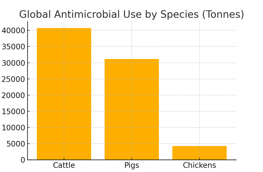
The study estimates that the global annual AMU for cattle, chickens, and pigs amounts to 76,060 tons of antimicrobial active ingredients. This is a significant revision from previous estimates due to a more detailed evaluation of animal weights and categories:
1. Cattle: 40,697 tons (53.5% of total AMU)
2. Pigs: 31,120 tons (40.9% of total AMU)
3. Chickens: 4,243 tons (5.6% of total AMU)
Figure 1: Distribution of global antimicrobial use among cattle, pigs, and chickens.
Methodology
The study utilizes the concept of Population Correction Units (PCU) to estimate antimicrobial usage, taking into account the weight and category of livestock at the time of treatment. This method differs from previous approaches that relied on live weight at slaughter, providing a more accurate representation of AMU.
The PCU is calculated by multiplying the number of animals by their average weight during treatment. This approach allows for differentiation by age and sex, which is particularly important for species like cattle and pigs.
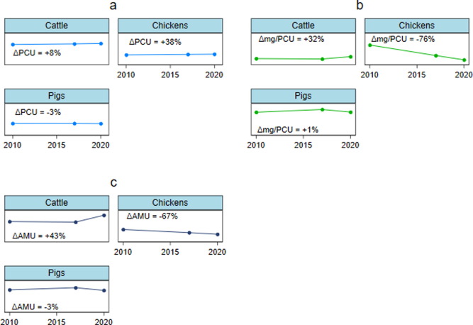
Figure 2: (a) Changes in global PCU (million tonnes), (b) changes in global antibiotic use in mg per PCU, and (c) changes in global AMU (thousand tonnes) for cattle, chickens, and pigs; between 2010 and 2020. Abbreviations: PCU = Population Correction Unit; AMU = Antibiotic Use.
Figure 2: (a) Changes in global PCU (million tonnes), (b) changes in global antibiotic use in mg per PCU, and (c) changes in global AMU (thousand tonnes) for cattle, chickens, and pigs; between 2010 and 2020. Abbreviations: PCU = Population Correction Unit; AMU = Antibiotic Use.
Study shows lower AMU than previous estimates
The study highlights a significant shift in AMU patterns, with chickens showing a remarkable decrease in antimicrobial use despite increased production. This is indicative of improved management and more responsible use of antibiotics in the poultry industry.
The lower AMU in cattle and pigs, compared to previous estimates, underscores the importance of considering animal age and weight at treatment. These findings align closely with World Organization for Animal Health (WOAH) estimates, validating the methodology.
However, the study also acknowledges limitations, including reliance on European standards for average weight at treatment, which may not reflect global variations. Additionally, the lack of comprehensive global data on veterinary antibiotics presents challenges in creating fully accurate estimates.
Corrected estimate highlights improved production advances
This study provides a revised and potentially more accurate estimate of global antimicrobial use in livestock. By accounting for the weight and treatment categories of animals, it offers insights that could guide policy and management practices to mitigate the spread of antimicrobial resistance.
The article also indicates that the industry may have over-estimated antimicrobial usage in livestock and, just as importantly, that antimicrobial use has been kept in check or even reduced, despite increases in farmed animal headcounts. The lower usage is likely due to regulatory oversight and improvements in alternative methods to control and mitigate health challenges.
Can phytogenics have a meaningful effect in coccidiosis control?
by Madalina Diaconu, Global Manager Gut Health, EW Nutrition
Coccidiosis, caused by Eimeria spp., is a major challenge in poultry production, leading to significant economic losses. Historically, control strategies have relied on chemical anticoccidials and ionophores. However, the emergence of drug-resistant Eimeria strains and consumer concerns about chemical residues necessitate alternative solutions. Phytogenics, especially tannins and saponins, offer promising natural solutions to be included in programs for coccidiosis control. More and more independent research highlights the potential of these natural compounds to enhance poultry health and productivity.
Efficacy of Tannins and Saponins in Coccidiosis Control
Phytogenics are plant-derived bioactive compounds known for their antimicrobial, antioxidant, and immunomodulatory properties. Among these, tannins and saponins have shown particular promise in supporting coccidiosis control.
The challenge: Preventing the spread of infections and mitigating subclinicial coccidiosis before it reaches this stage.
Tannins
Tannins are polyphenolic compounds found in various plants. They exhibit strong antimicrobial activity by binding to proteins and metal ions, disrupting microbial cell membranes, and inhibiting enzymatic activity.
Anticoccidial Activity: Tannins have been shown to interfere with the life cycle of Eimeria. Studies demonstrate that tannins can reduce oocyst shedding and intestinal lesion scores in infected birds (Abbas et al., 2017).
Immune Modulation: Tannins enhance immune responses by promoting the proliferation of lymphocytes and the production of antibodies, which help in the clearance of Eimeria infections (Redondo et al., 2021).
Saponins
Saponins are glycosides with surfactant properties, capable of lysing cell membranes of pathogens. They also stimulate immune responses, enhancing the host’s ability to fight infections.
Membrane Disruption: Saponins disrupt the cell membranes of Eimeria, leading to reduced parasite viability and replication (Githiori et al., 2004).
Immune Enhancement: Saponins stimulate the production of cytokines and enhance the activity of macrophages, improving the overall immune response against coccidiosis (Zhai et al., 2014).
Independent Research Evidences Phytogenics’s Role in Supporting Programs for Coccidiosis Control
Numerous studies have evaluated the efficacy of phytogenics in coccidiosis control. Here, we highlight key findings from peer-reviewed research:
Abbas et al. (2012): This study reviewed various botanicals and their effects on Eimeria species in poultry. The authors concluded that tannins and saponins significantly reduce oocyst shedding and lesion scores, comparable to conventional anticoccidials.
Allen et al. (1997): The authors investigated the use of dietary saponins in controlling Eimeria acervulina infections. The study found that saponin-treated birds exhibited lower oocyst counts and improved weight gain compared to untreated controls.
Masood et al. (2013): This study explored the role of natural antioxidants, including tannins, in controlling coccidiosis. The results indicated that tannins reduced oxidative stress and improved intestinal health, leading to better performance in broiler chickens.
Idris et al. (2017): The researchers assessed the potential of saponin-rich plant extracts against avian coccidiosis. The findings demonstrated significant reductions in oocyst output and lesion severity, highlighting the potential of saponins as effective anticoccidials.
Hailat et al. (2023): The researchers studied three phytogenic formulations against a control group with chemical drugs. The study concluded that phytogenic blends can be safely used as alternatives to the chemically synthesized drugs, either alone or in a shuttle program, for the control of poultry coccidiosis.
El-Shall et al. (2021): This review article highlights research findings on phytogenic compounds which showed preventive, therapeutic, or immuno-modulating effects against coccidiosis.
Despite initial skepticism, the growing body of evidence supports the efficacy of phytogenics in supporting coccidiosis control. Tannins and saponins, in particular, have shown significant potential in reducing parasite load, improving intestinal health, and enhancing immune responses. These natural compounds offer several advantages over traditional chemical treatments, including lower risk of resistance development and absence of harmful residues in meat products.
Challenges and Promises
While the efficacy of phytogenics is well-supported, challenges remain, especially with lower-quality products that may display variability in plant extract composition, in their standardization of doses, and in ensuring consistent quality. At the same time, these compounds are not silver bullets, and no producer should make unreasonable claims.
As far as the mode of action is concerned, the evidence is becoming clear: phytogenics, particularly tannins and saponins, are effective in mitigating gut health challenges and supporting bird performance when challenged. Their natural origin, coupled with potent antimicrobial and immunomodulatory properties, makes them suitable for sustainable poultry production. As the poultry industry seeks to reduce reliance on chemical drugs, phytogenics represent a viable and promising solution.
References
Abbas, R. Z., Iqbal, Z., Blake, D., Khan, M. N., & Saleemi, M. K. (2011). “Anticoccidial drug resistance in fowl coccidia: the state of play revisited”. World’s Poultry Science Journal, 67(2), 337-350. https://doi.org/10.1017/S004393391100033X
Allen, P. C., Danforth, H. D., & Levander, O. A. (1997). “Interaction of dietary flaxseed with coccidia infections in chickens”. Poultry Science, 76(6), 822-828. https://doi.org/10.1093/ps/76.6.822
El-Shall, N.A., El-Hack, M.E.A., et al. (2022). “Phytochemical control of poultry coccidiosis: a review”. Poultry Science, 101(1) 101542. https://doi.org/10.1016/j.psj.2021.101542
Idris, M., Abbas, R. Z., Masood, S., Rehman, T., Farooq, U., Babar, W., Hussain, R., Raza, A., & Riaz, U. (2017). “The potential of antioxidant rich essential oils against avian coccidiosis”. World’s Poultry Science Journal, 73(1), 89-104. https://doi.org/10.1017/S0043933916000787
Hailat, A.M., Abdelqader, A.M., & Gharaibeh, M.H. (2023). “Efficacy of Phyto-Genic Products to Control Field Coccidiosis in Broiler Chickens”. International Journal of Veterinary Science, 13(3), 266-272. https://doi.org/10.47278/journal.ijvs/2023.099
Masood, S., Abbas, R. Z., Iqbal, Z., Mansoor, M. K., Sindhu, Z. U. D., & Zia, M. A. (2013). “Role of natural antioxidants for the control of coccidiosis in poultry”. Pakistan Veterinary Journal, 33(4), 401-407.
Redondo, L. M., Chacana, P. A., Dominguez, J. E., & Miyakawa, M. E. (2021). “Perspectives in the use of tannins as alternative to antimicrobial growth promoter factors in poultry”. Frontiers in Microbiology, 12, 641949. https://doi.org/10.3389/fmicb.2021.641949
Zhai, H., Liu, H., Wang, S., Wu, J., & Kluenter, A. M. (2014). “Potential of essential oils for poultry and pigs”. Animal Nutrition, 2(4), 196-202. https://doi.org/10.1016/j.aninu.2016.12.004
Sustainable use of veterinary antimicrobials in Europe: EEA report
The European Environment Agency (EEA) has recently published a briefing detailing the impact of veterinary antimicrobials on Europe’s environment. Positive developments are to be applauded, however they do not tell the whole story.
The use of antimicrobials for farmed animals and in aquaculture decreased by around 28% between 2018 and 2022. Nonetheless, the rate of antimicrobial resistance continues to rise around the world, including as an important cause of death in the European Economic Area (the EEA includes all EU countries, as well as Norway, Lichtenstein, and Iceland). At present, antimicrobial-resistant infections are estimated to caused 35,000 deaths per year in the European Union. For reference, in the EU, traffic accidents cause around 20,000 deaths per year.
A large number of EU guidelines, policies, and regulations attempt to control and monitor the use of antimicrobials in food-producing animals. This makes the regulatory landscape somewhat confusing, especially that many implementation details are still left to the states.
Figure 1. Overview of the EU regulatory framework applicable to antimicrobials used in food-producing animals
One of the results of unequal implementation is that there is no standardized way of tracking the actual use of antimicrobials in food-producing animals. To collect the numbers, the EEA has used proxy numbers, especially antimicrobial sales data and self-reported data. With such broad strokes, it is to be expected that the actual figures might be higher.
Lower numbers…
According to the European Surveillance of Veterinary Antimicrobial Consumption (ESVAC) database, in the European Economic Area countries, plus Switzerland and the UK, total antimicrobial consumption for farmed animals and aquaculture was estimated at 73.9 mg/PCU* in 2022. This signifies a 30% reduction over 5 years.
In numbers: 4,458 tons of active substances were sold in one years for farmed animals & aquaculture.
*PCU represents a population correction unit (PCU). The PCU takes into account the population and relative weight of animals and “is used the normalize antimicrobial sales data for the size of the animal population that could potentially be treated with these substances. Using this methodology, 1 PCU corresponds to 1 kilogram of animal biomass”.
…But higher risks
In 2021, total antimicrobial consumption in humans – measured in 28 European countries – was estimated at 125.0 mg/kg. This number has unfortunately not gone down. What is worse: a much larger volume of antimicrobials is sold for food-producing animals than for human medicine. Which means that, relative to the total population, the impact of veterinary-use antimicrobials remains disproportionately large.
Moreover, two outliers (Poland and Lithuania) exhibited a worrying increasing trend, showing that no good development is irreversible. The EEA also highlights this danger, in the context of growing global consumption of animal protein. Increased demand “may put pressure on farmers to adopt intensive production practices that require increased use of antimicrobials”. The use of antimicrobials elsewhere in the world may lead to impacts in Europe, “not just by theoretically exposing consumers to antimicrobial residues but also by contributing to rising global rates of drug-resistant pathogens and infections”.
Figure 2. Sales by EU Member States in 2018 vs. 2022
Because of declining livestock populations in the last few years, while demand remained constant, EU-27 imports of animal products more than doubled between 2002 and 2022. There are no reliable global data on the veterinary use of antimicrobials, however it is generally believed that over 70% of antimicrobials sold globally may be used in for animal protein production.
Data collected by the World Organization for Animal Health (WOAH) from its member countries suggest that, between 2017-2019, “the use of antimicrobials in animals decreased by 25% in the Asia, Far East and Oceania regions, while it increased in Africa (+45%) and the Americas (+5%). Despite these partial improvements, a recent study forecasted that global use of antimicrobials in food-producing animals could rise by 8% in 2030, compared to 2020 levels (Mulchandani et al., 2023)”.
Quick summary
Reduction in Antimicrobial Use: There has been a significant reduction in the use of antimicrobials in farming and aquaculture across the EU. From 2018 to 2022, there was a decrease of approximately 28%, which aligns with the EU’s Farm to Fork strategy targeting a 50% reduction by 2030.
Antimicrobial Resistance (AMR): Despite the decrease in use, antimicrobial resistance remains a severe public health threat, causing an estimated 35,000 deaths annually in the European Economic Area. The resistance is attributed to the use of antimicrobials, which – as has been widely documented and discussed – can promote the evolution of resistant microorganisms.
Environmental Impact: The briefing underscores significant knowledge gaps in monitoring antimicrobial residues, resistant bacteria, and resistance genes in the environment. Improved surveillance could help identify pollution hotspots and assess the impact of reduction measures.
Regulatory and Policy Framework: The EU has implemented several policies to regulate the use of antimicrobials, including banning their use as growth promoters and setting stricter conditions for prescriptions. These measures are crucial for managing the risk of AMR.
Further efforts are needed to decrease the reliance on antimicrobials in food production. These include enhanced monitoring, promoting alternative practices in animal farming, and better animal welfare and biosecurity measures.
While improvements are clear and commendable in the EU-27 states, increased antimicrobial usage in some EU countries and in various areas around the world represent a significant concern.
For further details on the use of veterinary antimicrobials in Europe’s environment, you can refer to the EEA’s full report.
Organic acids can play a crucial role in zinc oxide replacement
Dr. Inge Heinzl, Editor EW Nutrition & Juan Antonio Mesonero Escuredo, GTM Swine/GPM Organic Acids EW Nutrition
The use of high levels of Zinc Oxide (ZnO) in the EU before 2022 was one of the most common methods to prevent postweaning diarrhea (PWD) in pig production. Pharmacologically high levels of ZnO (2000-3000 ppm) increase growth and reduce the incidence of enteric bacterial diseases such as post-weaning diarrhea (PWD)( Carlson et al., 1999; Hill et al., 2000; Hill et al., 2001; Poulsen & Larsen, 1995; De Mille et al., 2019).
However, ZnO showed adverse effects, such as the accumulation of heavy metal in the environment, the risk for antimicrobial resistance (AMR), and problems of mineral toxicity and adverse growth effects when feeding it longer than 28 days (Jensen et al., 2018; Cavaco et al., 2011; Vahjen, 2015; Romeo et al., 2014; Burrough et al., 2019). To replace ZnO in pig production, let us first look at its positive effects to know what we must compensate for.
ZnO has a multifactorial mode of action
ZnO shows several beneficial characteristics that positively influence gut health, the immune system, digestion, and, therefore, also overall health and growth performance.
Figure 1. Beneficial effects and ZnO mode of action in postweaning piglets
1. ZnO acts as an antimicrobial
Concerning the antimicrobial effects of ZnO, different possible modes of action are discussed:
ZnO in high dosages generates reactive oxygen species (ROS) that can damage the bacterial cell walls (Pasquet et al., 2014)
The death of the bacterial cell due to direct contact of the metallic Zn to the cell (Shearier et al., 2016)
Intrinsic antimicrobial properties of the ZnO2+ ions after dissociation. The uptake of zinc into cells is regulated by homeostasis. A concentration of the ZnO2+ ions higher than the optimal level of 10-7 to 10-5 M (depending on the microbial strain) allows the invasion of Zn2+ ions into the cell, and the zinc starts to be cytotoxic (Sugarman, 1983; Borovanský et al., 1989).
ZnO shows activity against, e.g., Staphylococcus aureus, Pseudomonas aeruginosa, E. coli,Streptococcus pyogenes, and other enterobacteria (Ann et al., 2014; Vahjen et al., 2016). However, Roselli et al. (2003) did not see a viability-decreasing effect of ZnO on ETEC.
2. ZnO modulates the immune system
Besides fighting pathogenic organisms as described in the previous chapter and supporting the immune system, ZnO is an essential trace element and has a vital role in the immune system. ZnO improves the innate immune response, increasing phagocytosis and oxidative bursts from macrophages and neutrophils. It also ameliorates the adaptative immune response by increasing the number of T lymphocytes (T cells) in general and regulatory T lymphocytes (T-regs) in particular. These cells control the immune response and inflammation (Kloubert et al., 2018). Macrophage capacity for phagocytosis (Ercan and Bor, 1991) and to kill parasites (Wirth et al., 1989), and also the killing activity of natural killer cells depends on Zn (Rolles et al., 2018). By reducing bacterial adhesion and blocking bacterial invasion, ZnO disburdens the immune system (Roselli et al., 2003).
ZnO reduces the expression of several proinflammatory cytokines induced by ETEC (Roselli et al., 2003). Several studies have also shown a modulation effect on intestinal inflammation, decreasing levels of IFN-γ, TNF-α, IL-1ß and IL-6, all pro-inflammatory, in piglets supplemented with ZnO (Zhu et al., 2017; Grilli et al., 2015).
3. ZnO improves digestion and promotes growth
Besides protecting young piglets against diarrhea, the goal is to make them grow optimally. For this target, an efficient digestion and a high absorption of nutrients is essential. Stimulating diverse pancreatic enzymes such as amylase, carboxypeptidase A, trypsin, chymotrypsin, and lipase increases digestibility (Hedemann et al., 2006; Pieper et al., 2015). However, Pieper et al. (2015) also showed that a long-term supply of very high dietary zinc triggers oxidative stress in the pancreas of piglets.
By stimulating the secretion of ghrelin at the stomach level and thereby promoting the release of insulin-like growth factor (IGF-1) and cholecystokinin (CCK), ZnO enhances muscle protein synthesis, cell proliferation, and feed intake (Yin et al., 2009; MacDonald et al., 2000)).
The result of improved digestion is increased body weight and average daily gain, which can be seen, e.g., in a study by Zhu et al. (2017).
4. ZnO protects the intestinal morphology
ZnO prevents the decrease of the trans-endothelial electrical resistance (TEER), usually occurring in the case of inflammation, by downregulating TNF-α and IFN-γ. TNF-α, as well as IFN-γ, increase the permeability of the epithelial tight junctions and, therefore, the intestinal barrier (Al-Sadi et al., 2009).
The enterotrophic and anti-apoptotic effect of ZnO is reflected by a higher number of proliferating and PCNA-positive cells and an increased mucosa surface in the ileum (higher villi, higher villi/crypt ratio)(Grilli et al., 2015). Zhu et al. (2017) also saw an increase in villus height in the duodenum and ileum and a decrease in crypt depth in the duodenum due to the application of 3000 mg of ZnO/kg. Additionally, they could notice a significant (P<0.05) upregulation of the mRNA expression of the zonula occludens-1 and occluding in the mucosa of the jejunum of weaned piglets.
In a trial conducted by Roselli et al. (2003), the supplementation of 0.2 mmol/L ZnO prevented the disruption of the membrane integrity when human Caco-2 enterocytes were challenged with ETEC.
5. ZnO acts antioxidant
The antioxidant effect of ZnO was shown in a study conducted by Zhu et al., 2017. They could demonstrate that the concentration of malondialdehyde (MDA), a marker for lipid peroxidation, decreased on day 14 or 28, and the total concentration of superoxide dismutase (SOD), comprising enzymes that transform harmful superoxide anions into hydrogen peroxide, increased on day 14 (P<0.05). Additionally, Zn is an essential ion for the catalytic action of these enzymes.
Which positive effects of ZnO can be covered by organic acids (OAs)?
1. OAs act antimicrobial
OAs, on the one hand, lower the pH in the gastrointestinal tract. Some pathogenic bacteria are susceptible to low pH. At a pH<5, the proliferation of, e.g., Salmonella, E. coli, and Clostridium is minimized. The good thing is that some beneficial bacteria, such as lactobacilli or bifidobacteria, survive as they are acid-tolerant. The lactobacilli, on their side, can produce hydrogen peroxide, which inhibits, e.g., Staphylococcus aureus or Pseudomonas spp. (Juven and Pierson, 1996).
Besides this more indirect mode of action, a more direct one is also possible: Owing to their lipophilic character, the undissociated form of OAs can pass the bacterial membrane (Partanen and Mroz, 1999). The lower the external pH, the more undissociated acid is available for invading the microbial cells. Inside the cell, the pH is higher than outside, and the OA dissociates. The release of hydrogen ions leads to a decrease in the internal pH of the cell and to a depressed cell metabolism. To get back to “normal conditions”, the cell expels protons. However, this is an energy-consuming process; longer exposure to OAs leads to cell death. The anion remaining in the cell, when removing the protons, disturbs the cell’s metabolic processes and participates in killing the bacterium.
These theoretical effects could be shown in a practical trial by Ahmed et al. (2014). He fed citric acid (0.5 %) and a blend of acidifiers composed of formic, propionic, lactic, and phosphoric acid + SiO2 (0.4 %) and saw a reduction in fecal counts of Salmonella and E. coli for both groups.
2. OAs modulate the immune system
The immune system is essential in the pig’s life, especially around weaning. Organic acids have been shown to support or stimulate the immune system. Citric acid (0.5%), as well as the blend of acidifiers mentioned before (Ahmed et al., 2014), significantly increased the level of serum IgG. IgG is part of the humoral immune system. They mark foreign substances to be eliminated by other defense systems.
Ren et al. (2019) could demonstrate a decrease in plasma tumor necrosis factor-α that regulates the activity of diverse immune cells. He also found lower interferon-γ and interleukin (Il)-1ß values in the OA group than in the control group after the challenge with ETEC. This trial shows that inflammatory response can be mitigated through the addition of organic acids.
3. OAs improve digestion and promote growth
In piglets, the acidity in the stomach is responsible for the activation and stimulation of certain enzymes. Additionally, it keeps the feed in the stomach for a longer time. Both effects lead to better digestion of the feed.
In the stomach, the conversion of pepsinogen to pepsin, which is responsible for protein digestion, is catalyzed under acid conditions (Sanny et al., 1975)group. Pepsin works optimally at two pH levels: pH 2 and pH 3.5 (Taylor, 1959). With increasing pH, the activity decreases; at pH 6, it stops. Therefore, a high pH can lead to poor digestion and undigested protein arriving in the intestine.
These final products of pepsin protein digestion are needed in the lower parts of the GIT to stimulate the secretion of pancreatic proteolytic enzymes. If they do not arrive, the enzymes are not activated, and the inadequate protein digestion continues. Additionally, gastric acid is the primary stimulant for bicarbonate secretion in the pancreas, neutralizing gastric acid and providing an optimal pH environment for the digestive enzymes working in the duodenum.
As already mentioned, the pH in the stomach influences the transport of digesta. The amount of digesta being transferred from the stomach to the small intestine is related to the acidity of the chyme leaving the stomach and arriving in the small intestine. Emptying of the stomach can only take place when the duodenal chyme can be neutralized by pancreatic or other secretions (Pohl et al., 2008); so, acid-sensitive receptors provide feedback regulation and a higher pH in the stomach leads to a faster transport of the digesta and a worse feed digestion.
4. OAs protect the intestinal morphology
Maintaining an intact gut mucosa with a high surface area is crucial for optimal nutrient absorption. Research suggests organic acids play a significant role in improving mucosal health:
Butyric acid promotes epithelial cell proliferation, as demonstrated in an in vitro pig hindgut mucosa study (Sakata et al., 1995). Fumaric acid, serving as an energy source, may locally enhance small intestinal mucosal growth, aiding in post-weaning epithelial cells’ recovery and increasing absorptive surface and digestive capacity (Blank et al., 1999). Sodium butyrate supplementation at low doses influences gastric morphology and function, thickening the stomach mucosa and enhancing mucosal maturation and differentiation (Mazzoni et al., 2008).
Studies show that organic acids affect gut morphology, with a mixture of short-chain and mid-chain fatty acids leading to longer villi (Ferrara et al., 2016) and Na-butyrate supplementation increasing crypt depth and villi length in the distal jejunum and ileum (Kotunia et al., 2004). However, the villi length and mucosa thickness in the duodenum were reduced. Dietary sodium butyrate has been linked to increased microvilli length and cecal crypt depth in pigs (Gálfi and Bokori, 1990).
5. OAs show antioxidant activity
The last characteristic, the antioxidant effect, cannot be provided at the same level as with ZnO; however, Zhang et al. (2019) attest to OAs a certain antioxidant activity. Oxalic, citric, acetic, malic, and succinic acids, which were extracted from Camellia oleifera, also showed good antioxidant activity in a trial conducted by Zhang et al. (2020).
Organic acids are an excellent tool to compensate for the ban on ZnO
The article shows that organic acids have similar positive effects as zinc oxide. They act antimicrobial, modulate the immune system, maintain the gut morphology, fight pathogenic microbes, and also act – slightly – antioxidant. Additionally, they have a significant advantage: they are not harmful to the environment. Organic acids used in the proper pH range and combination are good tools for replacing zinc oxide.
References on request
INFOGRAPHIC – Target measurements for water quality
Water is a main nutrient and carrier for vaccines, medicine – including antibiotics, but also for pathogens
Chemistry
pH and pKa
Acidity and dissociation index
Target: pH 3,5-3,8 Important for acids application (E.g. organic acids, etc), and ORP
Hardness
Content of Ca, sometimes plus Mg
Target: better TDS Important for acid binding capacity (ABC, buffer capacity)
Oxidation Reduction Potential (ORP)
Target: 650 mV>700 mV » reduces water intake Important for biocides application (E.g. chlorination)
Total Dissolved Solids (TDS)
Sum of dissolved salts, minerals, metals, carbonates, organics Target: 2000 ppm>3000 ppm » laxation Important for buffer capacity and ORP
Microbiology
Yeast
Target: < 5000 cfu/gr
Enterobacterias
Target: < 100 cfu/gr
Moulds
Target: < 100 cfu/gr
Nutritional considerations for immunity and gut health
Conference report
At the recent EW Nutrition Poultry Academy in Jakarta, Indonesia, Dr Steve Leeson, Professor Emeritus, University of Guelph, Canada, opened his presentation by stating that “it is obvious that any nutrient deficiency will impact bird health, but not so obvious is that nutrition per se can positively impact immunity and health in an otherwise healthy and high-producing bird.”
Modern high-performing broilers are characterized by extremely high feed intake. This puts a lot of stress on the physiology of the entire gastrointestinal tract, but particularly so on the absorptive epithelial cells of the small intestine. Any organism requires a nutrient source for survival and reproduction. Dr Leeson asked “can we significantly reduce nutrient supply to pathogens, while sustaining bird productivity?”
He reminded the audience that no cellular function comes for free: so there is always a “cost”. A general conclusion is that 10% of nutrients can be used for immune function during disease challenge, and always get priority. Therefore, you don’t want to overstimulate the immune system, which in extreme situations leads to an inflammatory response. In his presentation, Dr Leeson considered factors determining gut health and nutritional tools which are available to support gut health.
Gut microflora
Gut pathogens impact the bird and/or the consumer. Clostridia and E. coli are the major concerns regarding bird health and productivity, whereas Salmonella and Campylobacter are major pathogens important for human health.
The chick hatches with a gut virtually devoid of microbes, so early colonizers tend to predominate quite quickly. Microbial species present on the hatching tray, during delivery and during the first few days at the farm will likely dictate early gut colonization. In some instances, the chick’s microflora may be established by the time it gets to the farm, so the probiotic faces more of a challenge to establish itself as the predominant species.
Antibiotic alternatives
Gut villi development matures at around 10-15 days of age. The broiler pre-starter diet therefore is a target for feed additives that positively impact gut structure and development.
Among the short chain fatty acids, butyric acid is considered the prime energy source for enterocytes and it is also necessary for the correct development of the gut-associated lymphoid tissue (GALT). Butyric acid can also be added indirectly via fermentation of judicious levels of soluble fiber to encourage optimal gut villi development. Dr Leeson added that he is a big believer in butyric acid, encouraging a good gut structure at 10 days, which can be worth about 50 kcal.
Exogenous enzymes should also be considered in an attempt to maximize digestion and limit the flow of nutrients to the large intestine and ceca. Protease enzymes have great potential in this regard, since they allow nutritionists to reduce dietary crude protein and hopefully reduce the supply of nitrogen that fuels proteolytic Clostridia bacteria in the large intestine and ceca.
Amino acids, particularly threonine, play a critical role in the maintenance of intestinal mucosal integrity and barrier function, especially for mucin synthesis, which protects enterocytes from adherence by pathogenic bacteria, and from attack by endogenous enzymes and acids.
Polyunsaturated fatty acids (PUFAs) – Omega-3s and especially DHA from fish oil help to reduce inflammatory response (overstimulation). Omega-3s are poorly converted to DHA by the chicken, so conventional sources such as flax are of limited application for immunity.
Blood plasma from pigs or cattle is a complex spray-dried mixture of proteins and amino acids, many of which are immunoglobulins that “temper” the immune system, much like PUFAs.
Vitamins A, D, E and C have vital roles in the normal function of the immune system and have antioxidant capacity.
Certain complex carbohydrates, such as ß-glucans, influence gut health due to their fermentation, leading to the production of short-chain fatty acids, such as butyrate.
Antioxidants – to firstly control oxidation of fats and fat-soluble vitamins in feed, and secondly to optimize birds’ cellular oxidative capacity, to prevent cell damage, therefore maintaining healthy cellular and immune function.
Betaine increases intracellular water retention, reducing “dehydration” of microvilli and increasing their volume/surface area.
Fiber – moderate levels (1-2%) of soluble (fermentable) and insoluble fiber can be beneficial to early gut development by stimulating gizzard development and endogenous enzyme production.
Phytogenics are becoming very common in combination with acidifiers (upper tract) and probiotics. Essential oils are becoming more mainstream the more we know about them.
Recommendations for optimizing gut health and immunity
Fast growth rate and high egg output are negatively correlated with immune response. Consequently, nutrient-dense diets are not optimal for immunity. With bacteria, it’s a numbers game – but these numbers quickly multiply. The first 7 days are important, therefore probiotics must be established early. Consider the role of targeted feed additives, such as butyrate, phytogenics, antioxidants, PUFAs etc.
Also, maximize feed particle size – the limit is usually pellet quality. Mitigate nutrient transition at any diet change. Review the supply of trace minerals, as there is a trend to lower levels of organic minerals. With all the factors that weigh into production performance, any support that can be rallied through nutrition needs to be considered.
***
EW Nutrition’s Poultry Academy took place in Jakarta and Manila in early September 2023. Dr. Steve Leeson, an expert in Poultry Nutrition & Production with nearly 50 years’ experience in the industry, was the distinguished keynote speaker.
Dr. Leeson had his Ph.D. in Poultry Nutrition in 1974 from the University of Nottingham. Over a span of 38 years, he was a Professor in the Department of Animal &Poultry Science at the University of Guelph, Canada. Since 2014, he has been Professor Emeritus at the same University. As an eminent author, he has more than 400 papers in refereed journals and 6 books on various aspects of Poultry Nutrition & Management. He also won the American Feed Manufacturer’s Association Nutrition Research Award (1981), the Canadian Society of Animal Science Fellowship Award (2001), and Novus Lifetime Achievement Award in Poultry Nutrition (2011).
Salmonella in pigs: a threat for humans and a challenge for pig producers
By Dr. Inge Heinzl, Editor, EW Nutrition
Salmonellosis is third among foodborne diseases leading to death (Ferrari, 2019). More than 91,000 human cases of Salmonellosis are reported by the EU each year, generating overall costs of up to €3 billion a year (EFSA, 2023), 10-20% of which are attributed to pork consumption (Soumet, 2022). The annual costs arising from the resulting human health losses in 2010 were about €90 million (FCC Consortium, 2010). Take the example of Ireland, where a high prevalence of Salmonella in lymph nodes still shows a severe issue pre-slaughter and a big challenge for slaughterhouses to stick to the process hygiene requirements (Deane, 2022).
Several governments already have monitoring programs in place, and the farms are categorized according to the salmonella contamination of their pigs. In some countries, e.g., Denmark, an economic penalty of 2% of the carcass value must be paid if the farm has level 2 (intermediate seroprevalence) and 4-8% if the level is 3. Other countries, e.g., Germany, the UK, Ireland, or the Netherlands, use quality assurance schemes. The farmers can only sell their carcasses under this label if their farm has a certain level.
Let’s take a quick look at the genus of Salmonella
Salmonellas are rod-shaped gram-negative bacteria of the family of enterobacteria that use flagella for their movement. They were named after the American vet Daniel Elmer Salmon. The genus of Salmonella consists of two species (S. bongori and S. enterica with seven subspecies) with in total more than 2500 serovars (see Figure 1). The effects of the different serovars can range from asymptomatic carriage to severe invasive systemic disease (Gal-Mor, 2014). All Salmonella serovars generally can cause disease in humans; the rosa-marked ones already showed infections.
Figure 1: the genus of Salmonella with Salmonella serovars relevant for pigs (according to Bonardi, 2017: Salmonella in the pork production chain and its impact on human health in the European Union)
Within the group of Salmonella, some serovars can only reside in one or few species, e.g., S. enterica spp. enterica Serovar Dublin (S. Dublin) in bovines (Waldron, 2018) or S. Cholerasuis in pigs (Chiu, 2004). An infection in humans with these pathogens is often invasive and life-threatening (WHO, 2018). On the contrary, serovars like S. Typhimurium and S. Enteritidis are not host-specific and can cause disease in various species.
The serotypes S. Typhi and S. Paratyphi A, B, or C are highly adapted to humans and only for them pathogenic; they are responsible for the occurrence of typhus.
Transmission of Salmonella mostly happens via contaminated food
The way of transmission to humans depends on the serovar:
Human-specific and, therefore, only in humans and higher primates residing serovars S. Typhi and Paratyphi A, B, or C (typhoidal) are excreted via feces or urine. Therefore, any food or water contaminated with the feces or urine of infected people can transmit this disease (Government of South Australia, 2023). Typhoid and paratyphoid Salmonellosis occur endemic in developing countries with the lack of clean water and, therefore, inadequate hygiene (Gal-Mor, 2014).
Serovars which can cause disease in humans and animals (non-typhoidal), can be transmitted by
– animal products such as milk, eggs, meat
– contact with infected persons/animals (pigs, cows, pets, reptiles…) or
– other feces- or urine-contaminated products such as sprouts, vegetables, fruits….
Farm animals take salmonellas from their fellows, contaminated feed or water, rodents, or pests.
Symptoms of Salmonellosis can be severe
In the case of typhoid or paratyphoid Salmonellosis, the onset of illness is gradual. People can suffer from sustained high fever, unwellness, severe headache, and decreased appetite, but also from an enlarged spleen irritating the abdomen and dry cough.
A study conducted in Thailand with children suffering from enteric fever caused by the typhoid serovars S. Typhi and Paratyphi showed a sudden onset of fever and gastrointestinal issues (diarrhea), rose spots, bronchitis, and pneumonia (Thisyakorn et al., 1987)
The non-typhoid Salmonellosis is typically characterized by an acute onset of fever, nausea, abdominal pain with diarrhea, and sometimes vomiting (WHO, 2018). However, 5% of the persons – children with underlying conditions, e.g., babies, or people who have AIDS, malignancies, inflammatory bowel disease, gastrointestinal illness caused by non-typhoid serovars, and hemolytic anemia, or receiving an immunosuppressive therapy can be susceptible to bacteremia. Additionally, serovars like S. Cholerasuis or S. Dublin are apt to develop bacteremia by entering the bloodstream with little or no involvement of the gut (Chiu, 1999). In these cases, consequences can be septic arthritis, pneumonia, peritonitis, cutaneous abscess, mycotic aneurysm, and sometimes death (Chen et al., 2007; Chiu, 2004, Wang et al., 1996).
In pigs, S. Cholerasuis causes high fever, purple discolorations of the skin, and thereinafter diarrhea. The mortality rate in pigs suffering from this type of Salmonellosis is high. Barrows orally challenged with S. Typhimurium showed elevated rectal temperature by 12h, remaining elevated until the end of the study. Feed intake decreased with a peak at 48h after the challenge and remained up to 120h after the challenge. Daily gain reduced during the following two weeks after infection. A higher plasma cortisol level and a lower IGF-I level could also be noticed. All these effects indicate significant changes in the endocrine stress and the somatotropic axis, also without significant alterations in the systemic pro-inflammatory mediators (Balaji et al., 2000)
To protect humans, Salmonella in pork must be restraint
There are three main steps to keep the contamination of pork as low as possible:
Keeping Salmonella out of the pig farm
Minimizing spreading if Salmonella is already on the farm
Minimizing contamination in the slaughterhouse
1. How to keep Salmonella out of the pig farm?
To answer this question, we must look at how the pathogen can be transported to the farm. According to the Code of Practice for the Prevention and Control of Salmonella on Pig Farms (Ministry of Agriculture, Fisheries and Food and the Scottish Executive Rural Affairs Department), there are several possibilities to infiltrate the pathogen into the farm:
Diseased pigs or pigs which are ill but don’t show any symptoms
Feeding stuff or bedding contaminated with dung
Pets, rodents, wild birds, or animals
Farm personnel or visitors
Equipment or vehicles
Caution with purchased animals!
To minimize/prevent the entry of Salmonella into the livestock, bought-in animals must come from reputable breeding farms with a salmonella monitoring system in place. As possible carrier animals are more likely to excrete Salmonella when stressed; they should be kept in isolation after purchasing. Additionally, the animals must go through a disinfectant foot bath before entering the farm.
Keep rodents, wild animals, and vermin in check!
Generally, the production site must be kept clean and as unattractive as possible for all these animals. Rests of feed must be removed, and dead animals and afterbirths must be promptly and carefully disposed of. A well-planned baiting and trapping policy should be in place to effectively control rodents.
Only selected people should enter the hog houses
In any case, the number of persons entering the hog house must be kept as low as possible. Farmworkers should be trained in the principles of hygiene. They should wear adequate clothing (waterproof boots and protective overalls) that can be easily cleaned/laundered and disinfected. The clothes/shoes should always be used only at this site. Thorough hand washing and the disinfection of the boots when entering and leaving the pig unit are a must.
If visits are necessary, the visitors should take the same measures as the farm workers. And, of course, they should not have had contact with another pig farm during the last 48 hours.
Keep pens, farm equipment, and vehicles clean!
Farm equipment should not be shared with other farms. If this cannot be avoided, it must be cleaned and disinfected before re-entering the farm. Also, the vehicles for the transport of the animals must be cleaned and disinfected as soon as possible after usage, as contaminated transporters always pose the risk of infection.
Feed should be Salmonella-free!
To get high feed quality, the feed should be purchased from feed mills/sources with a well-functioning bacterial control to guarantee the absence of Salmonella. It is essential that birds, domestic and wild animals cannot enter the feed stores.
It is also advised to keep dry feed dry as possibly contaminating Salmonella can multiply in such humid conditions. Additionally, all feed bins and delivery pipes for dry and wet feed must be consciously cleaned, and the damp feed pipes also disinfected.
The change from pellets to mash could be helpful as the pellets facilitate Salmonella colonization by stimulating the secretion of mucins (Hedemann et al., 2005).
For sanitation of the feed, we offer organic acids (Acidomix product range) or mixtures of organic acids and formaldehyde in countries where formaldehyde products are allowed (Formycine) to decrease the pathogenic load of the feed materials. In vitro trials show the effectiveness of the products:
For the in vitro trial with Formycine, autoclaved feed samples were inoculated with Salmonella enteritidis serovar Typhimurium DSM 19587 strain to reach a Salmonella contamination of 106 CFU/g of feed. After incubating at room temperature for three hours, Formycine Liquido was added to the contaminated feed samples at 0, 500, 1000, and 2000 ppm. The control and inoculated feed samples were further incubated at room temperature, and Salmonella counts (CFU/g) were carried out at 24, 48, 72 hours and on day 15. The limit of Salmonella detection was set at 100 CFU/g (102). Results are shown in figure 2.
Fig. 2: Effect of treatment time and different inclusion levels of Formycine Liquido on the Salmonella count in feed
As important as uncontaminated feed is clean water for drinking. It can be achieved by taking the water from a main or a bacteriologically controlled water borehole. Regular cleaning/disinfection of the tanks, pipes, and drinkers is essential.
Bedding should be Salmonella-free
Straw material containing feces of other animals (rodents, pets) always carries the risk of Salmonella contamination. Also, wet or moldy bedding is not recommended because it is an additional challenge for the animal. To optimize the quality of bedding, the straw should be bought from reliable and as few as possible sources. The material must be stored dry and as far as practicable from the pig buildings (Ministry of Agriculture, Fisheries and Food & Scottish Executive Rural Affairs Department, 2000).
Vaccination is a beneficial measure
For the control of Salmonella in swine herds, vaccination is an effective tool. De Ridder et al. (2013) showed that an attenuated vaccine reduced the transmission of Salmonella Typhimurium in pigs. The vaccination with an attenuated S. Typhimurium strain, followed by a booster vaccination with inactivated S. Cholerasuis, showed better effects than an inactivated S. Cholerasuis vaccine alone (Alborali et al., 2017). Bearson et al. (2017) could delimitate transmission through less shedding and protect the animals against systemic disease.
To achieve the best effects, the producer must understand the diversity of Salmonella serovars to choose the most promising vaccination strategy (FSIS, 2023).
2. How to minimize the spreading of Salmonella on the farm?
If there are already cases of Salmonella on the farm, infected animals must be separated from the rest of the herd. Small batch sizes are beneficial, as well as not mixing different litters after weaning. If feasible, separate units for different production phases with an all-in/all-out system could break the reinfection cycle and help reduce Salmonella contamination on the farm. And also in this case, vaccination is helpful.
Salmonella doesn’t like acid conditions
An effective tool is acidifying the feed with organic acids, as Salmonella doesn’t like acid conditions. A trial was conducted with Acidomix AFG and Acidomix AFL to show their effects against Salmonella. For the test, 105 CFU/g of Salmonella enterica ser. Typhimurium was added to feed containing 1000 ppm, 2000 ppm, and 3000 ppm of Acidomix AFG or AFL. The stomach and intestine were simulated in vitro by adjusting the pH with HCl and NaHCO3 as follows:
Stomach 2.8
Intestine 6.8-7.0
After the respective incubation, the microorganisms were recovered from feed and plated on an appropriate medium for CFU counting. The results are shown in figures 3 and 4.
Combi
Figures 3 + 4: Effects of different concentrations of Acidomix AFG and Acidomix AFL against Salmonella enterica ser. Typhimurium in feed
Phytomolecules can support pigs against Salmonella
Plant compounds or phytomolecules can also be used against Salmonella in pigs. Some examples of phytomolecules to be used are Piperine, Allicin, Eugenol, and Carvacrol. Eugenol, e.g., increases the permeability of the Salmonella membrane, disrupts the cytoplasmic membrane, and inhibits the production of bacterial virulence factors (Keita et al., 2022; Mak et al., 2019). Thymol and Carvacrol interact with the cell membrane by H bonding, also resulting in a higher permeability.
An already published in vitro trial conducted with our product Ventar D also showed excellent effects against Salmonella while sparing the beneficial gut flora. A further trial once more demonstrated the susceptibility of Salmonella to Ventar D. It showed that Ventar D controls Salmonella by suppressing their motility and, at higher concentrations, inactivating the cells (see figures 5 + 6):
Figure 5: S. enterica motility test: on the left side – control; on the right side – motility medium containing.750 µg/mL of Ventar
Fig 6 . Disk diffusion assay employing S. enterica. upper left side – disk containing 10 µL of Ventar; upper right – 5 µL; lower left – control; lower right – 1µL.
In addition to the direct Salmonella-reducing effect, essential oils / secondary plant compounds / phytomolecules improve digestive enzyme activity and digestion, leading to increased nutrient absorption and better feed conversion (Windisch et al., 2008).
3. How can the farmer keep Salmonella contamination low in the slaughterhouse?
In general, the slaughterhouse personnel is responsible for adequate hygiene management to prevent contamination of carcasses and meat. However, also the farmer can make his contribution to maintain the risk of contamination in the slaughterhouse as low as possible. A study by Vieira-Pinto (2006) revealed that one Salmonella-positive pig can contaminate several other carcasses.
According to a trial conducted by Hurd et al. (2002), infection and, therefore, “contamination” of other pigs can rapidly occur, meaning that cross-contamination is a topic during transport to the slaughterhouse and in the lairages when the pigs come together with animals from other farms. The stress to which the pigs are exposed influences physiological and biochemical processes. The microbiome and animal’s immunity are affected, leading to higher excretion of Salmonella during transport and in the lairages. So, the animals should not be stressed during loading and unloading or transportation. The trailer poses a further risk of infection if it was not cleaned and disinfected before. So, reliable people who treat the animals well and keep their trailers clean should be chosen for transportation.
Pig producers are obliged to keep Salmonella in check – phytomolecules can help
At least in the EU, pig producers have the big duty to keep Salmonella low in their herds; otherwise, they will have financial losses. They are not only responsible for their farm, but also the slaughterhouses count on them. Besides the standard strict hygiene management and vaccination, farmers can use products provided by the industry to sanitize feed but also to support their animals directly with phytomolecules acting against pathogens and supporting gut health.
All these measures together should be a solution to the immense challenge of Salmonella, to protect people and prevent economic losses.
References:
Alborali, Giovanni Loris, Jessica Ruggeri, Michele Pesciaroli, Nicola Martinelli, Barbara Chirullo, Serena Ammendola, Andrea Battistoni, Maria Cristina Ossiprandi, Attilio Corradi, and Paolo Pasquali. “Prime-Boost Vaccination with Attenuated Salmonella Typhimurium Δznuabc and Inactivated Salmonella Choleraesuis Is Protective against Salmonella Choleraesuis Challenge Infection in Piglets.” BMC Veterinary Research 13, no. 1 (2017): 284. https://doi.org/10.1186/s12917-017-1202-5.
Balaji, R, K J Wright, C M Hill, S S Dritz, E L Knoppel, and J E Minton. “Acute Phase Responses of Pigs Challenged Orally with Salmonella Typhimurium.” Journal of Animal Science 78, no. 7 (2000): 1885. https://doi.org/10.2527/2000.7871885x.
Bearson, Bradley L, Shawn M. Bearson, Brian W Brunelle, Darrell O Bayles, In Soo Lee, and Jalusa D Kich. “Salmonella Diva Vaccine Reduces Disease, Colonization, and Shedding Due to Virulent S. Typhimurium Infection in Swine.” Journal of Medical Microbiology 66, no. 5 (2017): 651–61. https://doi.org/10.1099/jmm.0.000482.
Brenner Michael, G, M Cardoso, and S Schwarz. “Molecular Analysis of Salmonella Enterica Subsp. Enterica Serovar Agona Isolated from Slaughter Pigs.” Veterinary Microbiology 112, no. 1 (2006): 43–52. https://doi.org/10.1016/j.vetmic.2005.10.011.
Chen, P.-L., C.-M. Chang, C.-J. Wu, N.-Y. Ko, N.-Y. Lee, H.-C. Lee, H.-I. Shih, C.-C. Lee, R.-R. Wang, and W.-C. Ko. “Extraintestinal Focal Infections in Adults with Non-typhoid Salmonella Bacteraemia: Predisposing Factors and Clinical Outcome.” Journal of Internal Medicine 261, no. 1 (2007): 91–100. https://doi.org/10.1111/j.1365-2796.2006.01748.x.
Chiu, Cheng-Hsun, Lin-Hui Su, and Chishih Chu. “Salmonella EntericaSerotype Choleraesuis: Epidemiology, Pathogenesis, Clinical Disease, and Treatment.” Clinical Microbiology Reviews 17, no. 2 (2004): 311–22. https://doi.org/10.1128/cmr.17.2.311-322.2004.
De Ridder, L., D. Maes, J. Dewulf, F. Pasmans, F. Boyen, F. Haesebrouck, E. Méroc, P. Butaye, and Y. Van der Stede. “Evaluation of Three Intervention Strategies to Reduce the Transmission of Salmonella Typhimurium in Pigs.” The Veterinary Journal 197, no. 3 (2013): 613–18. https://doi.org/10.1016/j.tvjl.2013.03.026.
Deane, Annette, Declan Murphy, Finola C. Leonard, William Byrne, Tracey Clegg, Gillian Madigan, Margaret Griffin, John Egan, and Deirdre M. Prendergast. “Prevalence of Salmonella spp. in Slaughter Pigs and Carcasses in Irish Abattoirs and Their Antimicrobial Resistance.” Irish Veterinary Journal 75, no. 1 (2022). https://doi.org/10.1186/s13620-022-00211-y.
Edel, W., M. Schothorst, P. A. Guinée, and E. H. Kampelmacher. “Effect of Feeding Pellets on the Prevention and Sanitation of Salmonella Infections in Fattening Pigs1.” Zentralblatt für Veterinärmedizin Reihe B 17, no. 7 (2010): 730–38. https://doi.org/10.1111/j.1439-0450.1970.tb01571.x.
EFSA. “Salmonella.” European Food Safety Authority. Accessed August 7, 2023. https://www.efsa.europa.eu/en/topics/topic/salmonella.
Elbediwi, Mohammed, Daiwei Shi, Silpak Biswas, Xuebin Xu, and Min Yue. “Changing Patterns of Salmonella Enterica Serovar Rissen from Humans, Food Animals, and Animal-Derived Foods in China, 1995–2019.” Frontiers in Microbiology 12 (2021). https://doi.org/10.3389/fmicb.2021.702909.
Elnekave, Ehud, Samuel Hong, Alison E Mather, Dave Boxrud, Angela J Taylor, Victoria Lappi, Timothy J Johnson, et al. “Salmonella Enterica Serotype 4,[5],12:I:- In Swine in the United States Midwest: An Emerging Multidrug-Resistant Clade.” Clinical Infectious Diseases 66, no. 6 (2018): 877–85. https://doi.org/10.1093/cid/cix909.
Ferrari, Rafaela G., Denes K. Rosario, Adelino Cunha-Neto, Sérgio B. Mano, Eduardo E. Figueiredo, and Carlos A. Conte-Junior. “Worldwide Epidemiology of Salmonella serovars in Animal-Based Foods: A Meta-Analysis.” Applied and Environmental Microbiology 85, no. 14 (2019). https://doi.org/10.1128/aem.00591-19.
“FSIS Guideline to Control Salmonella in Swine Slaughter and Pork Processing Establishments.” FSIS Guideline to Control Salmonella in Swine Slaughter and Pork Processing Establishments | Food Safety and Inspection Service. Accessed August 14, 2023. https://www.fsis.usda.gov/guidelines/2023-0003.
Gal-Mor, Ohad, Erin C. Boyle, and Guntram A. Grassl. “Same Species, Different Diseases: How and Why Typhoidal and Non-Typhoidal Salmonella Enterica Serovars Differ.” Frontiers in Microbiology 5 (2014). https://doi.org/10.3389/fmicb.2014.00391.
González-Santamarina, Belén, Silvia García-Soto, Helmut Hotzel, Diana Meemken, Reinhard Fries, and Herbert Tomaso. “Salmonella Derby: A Comparative Genomic Analysis of Strains from Germany.” Frontiers in Microbiology 12 (2021). https://doi.org/10.3389/fmicb.2021.591929.
Government of South Australia. Typhoid and paratyphoid – including symptoms, treatment, and prevention, April 3, 2022. https://www.sahealth.sa.gov.au/wps/wcm/connect/public+content/sa+health+internet/conditions/infectious+diseases/typhoid+and+paratyphoid/typhoid+and+paratyphoid+-+including+symptoms+treatment+and+prevention.
Hauser, Elisabeth, Erhard Tietze, Reiner Helmuth, Ernst Junker, Kathrin Blank, Rita Prager, Wolfgang Rabsch, Bernd Appel, Angelika Fruth, and Burkhard Malorny. “Pork Contaminated with SalmonellaEnterica Serovar 4,[5],12:I:−, an Emerging Health Risk for Humans.” Applied and Environmental Microbiology 76, no. 14 (2010): 4601–10. https://doi.org/10.1128/aem.02991-09.
Health and Wellbeing; address=11 Hindmarsh Square, Adelaide scheme=AGLSTERMS.AglsAgent; corporateName=Department for. “Sa Health.” Typhoid and paratyphoid – including symptoms, treatment, and prevention, April 3, 2022. https://www.sahealth.sa.gov.au/wps/wcm/connect/public+content/sa+health+internet/conditions/infectious+diseases/typhoid+and+paratyphoid/typhoid+and+paratyphoid+-+including+symptoms+treatment+and+prevention.
Hedemann, M. S., L. L. Mikkelsen, P. J. Naughton, and B. B. Jensen. “Effect of Feed Particle Size and Feed Processing on Morphological Characteristics in the Small and Large Intestine of Pigs and on Adhesion of Salmonella Enterica Serovar Typhimurium DT12 in the Ileum in Vitro1.” Journal of Animal Science 83, no. 7 (2005): 1554–62. https://doi.org/10.2527/2005.8371554x.
Hendriksen, Susan W.M., Karin Orsel, Jaap A. Wagenaar, Angelika Miko, and Engeline van Duijkeren. “Animal-to-Human Transmission ofSalmonellaTyphimurium DT104A Variant.” Emerging Infectious Diseases 10, no. 12 (2004): 2225–27. https://doi.org/10.3201/eid1012.040286.
Keita, Kadiatou, Charles Darkoh, and Florence Okafor. “Secondary Plant Metabolites as Potent Drug Candidates against Antimicrobial-Resistant Pathogens.” SN Applied Sciences 4, no. 8 (2022). https://doi.org/10.1007/s42452-022-05084-y.
Ministry of Agriculture, Fisheries and Food, and Scottish Executive Rural Affairs Department. “Salmonella on Pig Farms – Code of Practice for the Prevention and Control Of.” ReadkonG.com, 2000. https://www.readkong.com/page/code-of-practice-for-the-prevention-and-control-of-5160969.
Morrow, W.E. Morgan, and Julie Funk. Ms. Salmonella as a Foodborne Pathogen in Pork. North Carolina State University Animal Science, n.d.
Soumet, C., A. Kerouanton, A. Bridier, N. Rose, M. Denis, I. Attig, N. Haddache, and C. Fablet. Report, Salmonella excretion level in pig farms and impact of quaternary ammonium compounds based disinfectants on Escherichia coli antibiotic resistance § (2022).
Thisyakorn, Usa. “Typhoid and Paratyphoid Fever in 192 Hospitalized Children in Thailand.” Archives of Pediatrics & Adolescent Medicine 141, no. 8 (1987): 862. https://doi.org/10.1001/archpedi.1987.04460080048025.
Ung, Aymeric, Amrish Y. Baidjoe, Dieter Van Cauteren, Nizar Fawal, Laetitia Fabre, Caroline Guerrisi, Kostas Danis, et al. “Disentangling a Complex Nationwide Salmonella Dublin Outbreak Associated with Raw-Milk Cheese Consumption, France, 2015 to 2016.” Eurosurveillance 24, no. 3 (2019). https://doi.org/10.2807/1560-7917.es.2019.24.3.1700703.
Vieira-Pinto, M, R Tenreiro, and C Martins. “Unveiling Contamination Sources and Dissemination Routes of Salmonella Sp. in Pigs at a Portuguese Slaughterhouse through Macrorestriction Profiling by Pulsed-Field Gel Electrophoresis.” International Journal of Food Microbiology 110, no. 1 (2006): 77–84. https://doi.org/10.1016/j.ijfoodmicro.2006.01.046.
Waldron, P. “Keeping Cows and Humans Safe from Salmonella Dublin.” Cornell University College of Veterinary Medicine, December 25, 2018. https://www.vet.cornell.edu/news/20181218/keeping-cows-and-humans-safe-salmonella-dublin.
Wang, J.-H., Y.-C. Liu, M.-Y. Yen, J.-H. Wang, Y.-S. Chen, S.-R. Wann, and D.-L. Cheng. “Mycotic Aneurysm Due to Non-Typhi Salmonella: Report of 16 Cases.” Clinical Infectious Diseases 23, no. 4 (1996): 743–47. https://doi.org/10.1093/clinids/23.4.743.
WHO. “Salmonella (Non-Typhoidal).” World Health Organization, February 20, 2018. https://www.who.int/news-room/fact-sheets/detail/salmonella-(non-typhoidal).
Windisch, W., K. Schedle, C. Plitzner, and A. Kroismayr. “Use of Phytogenic Products as Feed Additives for Swine and Poultry1.” Journal of Animal Science 86, no. suppl_14 (2008). https://doi.org/10.2527/jas.2007-0459.
Windisch, W., K. Schedle, C. Plitzner, and A. Kroismayr. “Use of Phytogenic Products as Feed Additives for Swine and Poultry1.” Journal of Animal Science 86, no. suppl_14 (2008). https://doi.org/10.2527/jas.2007-0459.
Minimizing Collateral Effects of Antibiotic Administration in Swine Farms: A Balancing Act
By Dr Merideth Parke BVSc, Regional Technical Manager Swine, EW Nutrition
We care for our animals, and antibiotics are a crucial component in the management of disease due to susceptible pathogens, supporting animal health and welfare. However, the administration of antibiotics in pig farming has become a common practice to prevent bacterial infections, reduce economic losses, and increase productivity.
All antibiotic applications have collateral consequences of significance, bringing a deeper consideration to their non-essential application. This article aims to challenge the choice to administer antibiotics by exploring the broader impact that antibiotics have on animal and human health, economies, and the environment.
Antibiotics disrupt microbial communities
Antibiotics do not specifically target pathogenic bacteria. By impacting beneficial microorganisms, they disrupt the natural balance of microbial communities within animals. They reduce the microbiota diversity and abundance of all susceptible bacteria – beneficial and pathogenic ones… many of which play crucial roles in digestion, brain function, the immune system, and respiratory and overall health. Resulting microbiota imbalances may present themselves in animals showing health performance changes associated with non-target systems, including the nasal, respiratory, or gut microbiome10, 9, 16. The gut-respiratory microbiome axis is well-established in mammals. Gut microbiota health, diversity, and nutrient supply directly impact respiratory health and function15. In pigs specifically, the modulation of the gut microbiome is being considered as an additional tool in the control of respiratory diseases such as PRRS due to the link between the digestion of nutrients, systemic immunity, and response to pulmonary infections12.
The collateral effect of antibiotic administration disrupting not only the microbial communities throughout the animal but also linked body systems needs to be considered significant in the context of optimal animal health, welfare, and productivity.
Antibiotic use can lead to the release of toxins
The consideration of the pathogenesis of individual bacteria is critical to mitigate potential for direct collateral effects associated with antibiotic administration. For example, in cases of toxin producing bacteria, when animals are medicated either orally or parenterally, mortality may increase due to the associated release of toxins when large numbers of toxin producing bacteria are killed quickly3.
Modulation of the brain function can be critical
Numerous animal studies have investigated the modulatory role of intestinal microbes on the gut-brain axis. One identified mechanism seen with antibiotic-induced changes in fecal microbiota is the decreased concentrations of hypothalamic neurotransmitter precursors, 5-hydroxytryptamine (serotonin), and dopamine6. Neurotransmitters are essential for communication between the nerve cells. Animals with oral antibiotic-induced microbiota depletion have been shown to experience changes in brain function, such as spatial memory deficits and depressive-like behaviors.
Processing of waste materials can be impacted
Anaerobic treatment technology is well accepted as a feasible management process for swine farm wastewater due to its relatively low cost with the benefit of bioenergy production. Additionally, the much smaller volume of sludge remaining after anaerobic processing further eases the safe disposal and decreases the risk associated with the disposal of swine waste containing residual antibiotics5.
The excretion of antibiotics in animal waste, and the resulting presence of antibiotics in wastewater, can impact the success of anaerobic treatment technologies, which already could be demonstrated by several studies8, 13. The degree to which antibiotics affect this process will vary by type, combination, and concentration. Furthermore, the presence of antibiotics within the anaerobic system may result in a population shift towards less sensitive microbes or the development of strains with antibiotic-resistant genes1, 14.
Antibiotics can be transferred to the human food chain
Regulatory authorities specify detailed withdrawal periods after antibiotic treatment. However, residues of antibiotics and their metabolites may persist in animal tissues, such as meat and milk, even after this period. These residues can enter the human food chain if not adequately monitored and controlled.
Prolonged exposure to low levels of antibiotics through the consumption of animal products may contribute to the emergence of antibiotic-resistant bacteria in humans, posing a significant public health risk.
Contamination of the environment
As already mentioned before, the administration of antibiotics to livestock can result in the release of these compounds into the environment. Antibiotics can enter the soil, waterways, and surrounding ecosystems through excretions from treated animals, inappropriate disposal of manure, and runoff from agricultural fields. Once in the environment, antibiotics can contribute to the selection and spread of antibiotic-resistant bacteria in natural bacterial communities. This contamination poses a potential risk to wildlife, including birds, fish, and other aquatic organisms, as well as the broader ecological balance of affected ecosystems.
Every use of antibiotics can create resistance
One of the widely researched concerns associated with antibiotic use in livestock is the development of antibiotic resistance. The development of AMR does not require prolonged antibiotic use and, along with other collateral effects, also occurs when antibiotics are used within recommended therapeutic or preventive applications.
Gene mutations can supply bacteria with abilities that make them resistant to certain antibiotics (e.g., a mechanism to destroy or discharge the antibiotic). This resistance can be transferred to other microorganisms, as seen with the effect of carbadox on Escherichia coli7 and Salmonella enterica2 and the carbadox and metronidazole effect on Brachyspira hyodysenteriae16. Additionally, there is an indication that the zinc resistance of Staphylococcus of animal origin is associated with the methicillin resistance coming from humans4.
Consequently, the effectiveness of antibiotics in treating infections in target animals becomes compromised, and the risk of exposure to resistant pathogens for in-contact animals and across species increases, including humans.
Alternative solutions are available
To successfully minimize the collateral effects of antibiotic administration in livestock, a unified strategy with support from all stakeholders in the production system is essential. The European Innovation Partnership – Agriculture11 concisely summarizes such a process as requiring…
Changing human mindsets and habits: this is the first and defining step to successful antimicrobial reduction
Improving pig health and welfare: Prevention of disease with optimal husbandry, hygiene, biosecurity, vaccination programs, and nutritional support.
Effective antibiotic alternatives: for this purpose, phytomolecules, pro/pre-biotics, organic acids, and immunoglobulins are considerations.
In general, implementing responsible antibiotic stewardship practices is paramount. This includes limiting antibiotic use to the treatment of diagnosed infections with an effective antibiotic, and eliminating their use as growth promotors or for prophylactic purposes.
Keeping the balance is of crucial importance
While antibiotics play a crucial role in ensuring the health and welfare of livestock, their extensive administration in the agricultural industry has collateral effects that cannot be ignored. The development of antibiotic resistance, environmental contamination, disruption of microbial communities, and the potential transfer of antibiotic residues to food pose significant challenges.
Adopting responsible antibiotic stewardship practices, including veterinary oversight, disease prevention programs, optimal animal husbandry practices, and alternatives to antibiotics, can strike a balance between animal health, efficient productive performance, and environmental and human health concerns.
The collaboration of stakeholders, including farmers, veterinarians, policymakers, industry and consumers, is essential in implementing and supporting these measures to create a sustainable and resilient livestock industry.
References
Angenent, Largus T., Margit Mau, Usha George, James A. Zahn, and Lutgarde Raskin. “Effect of the Presence of the Antimicrobial Tylosin in Swine Waste on Anaerobic Treatment.” Water Research 42, no. 10–11 (2008): 2377–84. https://doi.org/10.1016/j.watres.2008.01.005.
Bearson, Bradley L., Heather K. Allen, Brian W. Brunelle, In Soo Lee, Sherwood R. Casjens, and Thaddeus B. Stanton. “The Agricultural Antibiotic Carbadox Induces Phage-Mediated Gene Transfer in Salmonella.” Frontiers in Microbiology 5 (2014). https://doi.org/10.3389/fmicb.2014.00052.
Castillofollow, Manuel Toledo, Rocío García Espejofollow, Alejandro Martínez Molinafollow, María Elena Goyena Salgadofollow, José Manuel Pintofollow, Ángela Gallardo Marínfollow, M. Toledo, et al. “Clinical Case: Edema Disease – the More I Medicate, the More Pigs Die!” $this->url_servidor, October 15, 2021. https://www.pig333.com/articles/edema-disease-the-more-i-medicate-the-more-pigs-die_17660/.
Cavaco, Lina M., Henrik Hasman, Frank M. Aarestrup, Members of MRSA-CG:, Jaap A. Wagenaar, Haitske Graveland, Kees Veldman, et al. “Zinc Resistance of Staphylococcus Aureus of Animal Origin Is Strongly Associated with Methicillin Resistance.” Veterinary Microbiology 150, no. 3–4 (2011): 344–48. https://doi.org/10.1016/j.vetmic.2011.02.014.
Cheng, D.L., H.H. Ngo, W.S. Guo, S.W. Chang, D.D. Nguyen, S. Mathava Kumar, B. Du, Q. Wei, and D. Wei. “Problematic Effects of Antibiotics on Anaerobic Treatment of Swine Wastewater.” Bioresource Technology 263 (2018): 642–53. https://doi.org/10.1016/j.biortech.2018.05.010.
Köhler, Bernd, Helge Karch, and Herbert Schmidt. “Antibacterials That Are Used as Growth Promoters in Animal Husbandry Can Affect the Release of Shiga-Toxin-2-Converting Bacteriophages and Shiga Toxin 2 from Escherichia Coli Strains.” Microbiology 146, no. 5 (2000): 1085–90. https://doi.org/10.1099/00221287-146-5-1085.
Loftin, Keith A., Cynthia Henny, Craig D. Adams, Rao Surampali, and Melanie R. Mormile. “Inhibition of Microbial Metabolism in Anaerobic Lagoons by Selected Sulfonamides, Tetracyclines, Lincomycin, and Tylosin Tartrate.” Environmental Toxicology and Chemistry 24, no. 4 (2005): 782–88. https://doi.org/10.1897/04-093r.1.
Looft, Torey, Heather K Allen, Brandi L Cantarel, Uri Y Levine, Darrell O Bayles, David P Alt, Bernard Henrissat, and Thaddeus B Stanton. “Bacteria, Phages and Pigs: The Effects of in-Feed Antibiotics on the Microbiome at Different Gut Locations.” The ISME Journal 8, no. 8 (2014a): 1566–76. https://doi.org/10.1038/ismej.2014.12.
Looft, Torey, Heather K. Allen, Thomas A. Casey, David P. Alt, and Thaddeus B. Stanton. “Carbadox Has Both Temporary and Lasting Effects on the Swine Gut Microbiota.” Frontiers in Microbiology 5 (2014b). https://doi.org/10.3389/fmicb.2014.00276.
Nasralla, Meisoon. “EIP-Agri Concept.” EIP-AGRI – European Commission, September 11, 2017. https://ec.europa.eu/eip/agriculture/en/eip-agri-concept.html.
Niederwerder, Megan C. “Role of the Microbiome in Swine Respiratory Disease.” Veterinary Microbiology 209 (2017): 97–106. https://doi.org/10.1016/j.vetmic.2017.02.017.
Poels, J., P. Van Assche, and W. Verstraete. “Effects of Disinfectants and Antibiotics on the Anaerobic Digestion of Piggery Waste.” Agricultural Wastes 9, no. 4 (1984): 239–47. https://doi.org/10.1016/0141-4607(84)90083-0.
Shimada, Toshio, Julie L. Zilles, Eberhard Morgenroth, and Lutgarde Raskin. “Inhibitory Effects of the Macrolide Antimicrobial Tylosin on Anaerobic Treatment.” Biotechnology and Bioengineering 101, no. 1 (2008): 73–82. https://doi.org/10.1002/bit.21864.
Sikder, Md. Al, Ridwan B. Rashid, Tufael Ahmed, Ismail Sebina, Daniel R. Howard, Md. Ashik Ullah, Muhammed Mahfuzur Rahman, et al. “Maternal Diet Modulates the Infant Microbiome and Intestinal Flt3l Necessary for Dendritic Cell Development and Immunity to Respiratory Infection.” Immunity 56, no. 5 (May 9, 2023): 1098–1114. https://doi.org/10.1016/j.immuni.2023.03.002.
Slifierz, Mackenzie Jonathan. “The Effects of Zinc Therapy on the Co-Selection of Methicillin-Resistance in Livestock-Associated Staphylococcus Aureus and the Bacterial Ecology of the Porcine Microbiota,” 2016.
Stanton, Thaddeus B., Samuel B. Humphrey, Vijay K. Sharma, and Richard L. Zuerner. “Collateral Effects of Antibiotics: Carbadox and Metronidazole Induce VSH-1 and Facilitate Gene Transfer among Brachyspira Hyodysenteriae” Applied and Environmental Microbiology 74, no. 10 (2008): 2950–56. https://doi.org/10.1128/aem.00189-08.
The future of coccidiosis control
By Madalina Diaconu, Product Manager Pretect D, EW Nutrition and Twan van Gerwe, Ph.D., Technical Director, EW Nutrition
With costs of over 14 billion USD per year (Blake, 2020), coccidiosis is one of the most devastating enteric challenges in the poultry industry. With regard to costs, subclinical forms of coccidiosis account for the majority of production losses, as damage to intestinal cells results in lower body weight, higher feed conversion rates, lack of flock uniformity, and failures in skin pigmentation. This challenge can only be tackled, if we understand the basics of coccidiosis control in poultry and what options producers have to manage coccidiosis risks.
Current strategies show weak points
Good farm management, litter management, and coccidiosis control programs such as shuttle and rotation programs form the basis for preventing clinical coccidiosis. More successful strategies include disease monitoring, strategic use of coccidiostats, and increasingly coccidiosis vaccines. However, the intrinsic properties of coccidia make these parasites often frustrating to control. Acquired resistance to available coccidiostats is the most difficult and challenging factor to overcome.
Optimally, coccidiosis control programs are developed based on the farm history and the severity of infection. The coccidiostats traditionally used were chemicals and ionophores, with ionophores being polyether antibiotics. To prevent the development of resistance, the coccidiostats were used in shuttle or rotation programs, at which in the rotation program, the anticoccidial changes from flock to flock, and in the shuttle program within one production cycle (Chapman, 1997).
The control strategies, however, are not 100% effective. The reason for that is a lack of diversity in available drug molecules and the overuse of some molecules within programs. An additional lack of sufficient coccidiosis monitoring and rigorous financial optimization often leads to cost-saving but only marginally effective solutions. At first glance, they seem effective, but in reality, they promote resistance, the development of subclinical coccidiosis, expressed in a worsened feed conversion rate, and possibly also clinical coccidiosis.
Market requests and regulations drive coccidiosis control strategies
Changing coccidiosis control strategies has two main drivers: the global interest in mitigating antimicrobial resistance and the consumer’s demand for antibiotic-free meat production.
Authorities have left ionophores untouched
Already in the late 1990s, due to the fear of growing antimicrobial resistance, the EU withdrew the authorization for Avoparcin, Bacitracin zinc, Spiramycin, Virginiamycin, and Tylosin phosphate, typical growth promoters, to “help decrease resistance to antibiotics used in medical therapy”. However, ionophores, being also antibiotics, were left untouched: The regulation (EC) No 1831/2003 [13]of the European Parliament and the Council of 22 September 2003 clearly distinguished between coccidiostats and antibiotic growth promoters. Unlike the antibiotic growth promoters, whose primary action site is the gut microflora, coccidiostats only have a secondary and residual activity against the gut microflora. Furthermore, the Commission declared in 2022 that the use of coccidiostats would not presently be ruled out “even if of antibiotic origin” (MEMO/02/66, 2022) as “hygienic precautions and adaptive husbandry measures are not sufficient to keep poultry free of coccidiosis” and that “modern poultry husbandry is currently only practicable if coccidiosis can be prevented by inhibiting or killing parasites during their development”. In other words, the Commission acknowledged that ionophores were only still authorized because it believed there were no other means of controlling coccidiosis in profitable poultry production.
Consumer trends drove research on natural solutions
Due to consumers’ demand for antibiotic-reduced or, even better, antibiotic-free meat production, intensified industrial research to fight coccidiosis with natural solutions has shown success. Knowledge, research, and technological developments are now at the stage of offering solutions that can be an effective part of the coccidia control program and open up opportunities to make poultry production even more sustainable by reducing drug dependency.
Producers from other countries have already reacted. Different from the handling of ionophores regime in the EU, where they are allowed as feed additives, in the United States, coccidiostats belonging to the polyether-ionophore class are not permitted in NAE (No Antibiotics Ever) and RWE (Raised Without Antibiotics) programs. Instead of using ionophores, coccidiosis is controlled with a veterinary-led combination of live vaccines, synthetic compounds, phytomolecules, and farm management. This approach can be successful, as demonstrated by the fact that over 50% of broiler meat production in the US is NAE. Another example is Australia, where the two leading retail store chains also exclude chemical coccidiostats from broiler production. In certain European countries, e.g., Norway, the focus is increasingly on banning ionophores.
The transition to natural solutions needs knowledge and finesse
In the beginning, the transition from conventional to NAE production can be difficult. There is the possibility to leave out the ionophores and manage the control program only with chemicals of different modes of action. More effective, however, is a combination of vaccination and chemicals (bio-shuttle program) or the combination of phytomolecules with vaccination and/or chemicals (Gaydos, 2022).
Coccidiosis vaccination essentials
When it is decided that natural solutions shall be used to control coccidiosis, some things about vaccination must be known:
There are different strains of vaccines, natural ones selected from the field and attenuated strains. The formers show medium pathogenicity and enable a controlled infection of the flock. The latter, being early mature lower pathogenicity strains, usually cause only low or no post-vaccinal reactions.
A coccidiosis program that includes vaccination should cover the period from the hatchery till the end of the production cycle. Perfect application of the vaccines and effective recirculation of vaccine strains amongst the broilers are only two examples of preconditions that must be fulfilled for striking success and, therefore, early and homogenous immunity of the flock.
Perfect handling of the vaccines is of vital importance. For that purpose, the personnel conducting the vaccinations in the hatchery or on the farms must be trained. In some situations, consistent high-quality application at the farm has shown to be challenging. As a result, interest in vaccine application at the hatchery is growing.
Phytochemicals are a perfect tool to complement coccidiosis control programs
As the availability of vaccines is limited and the application costs are relatively high, the industry has been researching supportive measures or products and discovered phytochemicals as the best choice. Effective phytochemical substances have antimicrobial and antiparasitic properties and enhance protective immunity in poultry infected by coccidiosis. They can be used in rotation with vaccination, to curtail vaccination reactions of (non-attenuated) wild strain vaccines, or in combination with chemical coccidiostats in a shuttle program.
In a recent review paper (El-Shall et al., 2022), natural herbal products and their extracts have been described to effectively reduce oocyst output by inhibiting Eimeria species’ invasion, replication, and development in chicken gut tissues. Phenolic compounds in herbal extracts cause coccidia cell death and lower oocyst counts. Additionally, herbal additives offer benefits such as reducing intestinal lipid peroxidation, facilitating epithelial repair, and decreasing Eimeria-induced intestinal permeability.
Various phytochemical remedies are shown in this simplified adaptation of a table from El-Shall et al. (2022), indicating the effects exerted on poultry in connection to coccidia infection.
Bioactive compound
Effect
Saponins
Inhibition of coccidia:
By binding to membrane cholesterol, the saponins disturb the lipids in the parasite cell membrane. The impact on the enzymatic activity and metabolism leads to cell death, which then induces a toxic effect in mature enterocytes in the intestinal mucosa. As a result, sporozoite-infected cells are released before the protozoa reach the merozoite phase.Support for the chicken:
Saponins enhance non-specific immunity and increase productive performance (higher daily gain and improved FCR, lower mortality rate). They decrease fecal oocyst shedding and reduce ammonia production.
Tannins
Inhibition of coccidia:
Tannins penetrate the coccidia oocyst wall and inactivate the endogenous enzymes responsible for sporulation.Support for the chicken: Additionally, they enhance anticoccidial antibodies’ activity by increasing cellular and humoral immunity.
Flavonoids and terpenoids
Inhibition of coccidia:
They inhibit the invasion and replication of different species of coccidia.Support for the chicken: They bind to the mannose receptor on macrophages and stimulate them to produce inflammatory cytokines such as IL-1 through IL-6 and TNF. Higher weight gain and lower fecal oocyst output are an indication of suppression of coccidiosis.
Artemisinin
Inhibition of coccidia:
Its impact on calcium homeostasis compromises the oocyst wall formation and leads to a defective cell wall and, in the end, to the death of the oocyst. Enhancing the production of ROS directly inhibits sporulation and also wall formation and, therefore, affects the Eimeria life cycle.Support for the chicken: Reduction of oocyst shedding
Leaf powder of Artemisia annua
Support for the chicken: Protection from pathological symptoms and mortality associated with Eimeria tenella infection. Reduced lesion score and fecal oocyst output.
The leaf powder was more efficient than the essential oil, which could be due to a lack of Artemisinin in the oil, and to the greater antioxidant ability of A. annua leaves than the oil.
Phenols
Inhibition of coccidia:
Phenols change the cytoplasmic membrane’s permeability for cations (H+ and K+), impairing essential processes in the cell. The resulting leakage of cellular constituents leads to water unbalance, collapse of the membrane potential, inhibition of ATP synthesis, and, finally, cell death. Due to their toxic effect on the upper layer of mature enterocytes of the intestinal mucosa, they accelerate the natural renewal process, and, therefore, sporozoite-infected cells are shed before the coccidia reaches the merozoite phase.
Table 1: Bioactive compounds and their anticoccidial effect exerted in poultry
Consumers vote for natural – phytochemicals are the solution
Due to still rising antimicrobial resistance, consumers push for meat production without antimicrobial usage. Phytomolecules, as a natural solution, create opportunities to make poultry production more sustainable by reducing dependency on harmful drugs. With their advent, there is hope that antibiotic resistance can be held in check without affecting the profitability of poultry farming.
Fighting antimicrobial resistance with immunoglobulins
By Lea Poppe, Regional Technical Manager On-Farm Solutions Europe, and Dr. Inge Heinzl, Editor
One of the ten global public health threats is antimicrobial resistance (AMR). Jim O’Neill predicted 10 million people dying from AMR annually by 2050 (O’Neill, 2016). The following article will show the causes of antimicrobial resistance and how antibodies from the egg could help mitigate the problem of AMR.
Global problem of AMR results from the incorrect use of antimicrobials
Antimicrobial substances are used to prevent and cure diseases in humans, animals, and plants and include antibiotics, antivirals, antiparasitics, and antifungals. The use of these medicines does not always happen consciously, partially due to ignorance and partially for economic reasons.
There are various possibilities for the wrong therapy
The use of antibiotics against diseases that household remedies could cure. A recently published German study (Merle et al., 2023) confirmed the linear relationship between treatment frequency and resistant scores in calves younger than eight months.
The use of antibiotics against viral diseases: antibiotics only act against bacteria and not against viruses. Flu, e.g., is caused by a virus, but doctors often prescribe an antibiotic.
Using broad-spectrum antibiotics instead of determining an antibiogram and applying a specific antibiotic.
A too-long treatment with antimicrobials so that the microorganisms have the time to adapt. For a long time, the only mistake you could make was to stop the antibiotic therapy too early. Today, the motto is “as short as possible”.
Let’s take the example of neonatal calf diarrhea, one of the most common diseases with a high economic impact. Calf diarrhea can be caused by a wide range of bacteria, viruses, or parasites. This infectious form can be a complication of non-infectious diarrhea caused by dietary, psychological, and environmental stress (Uetake, 2012). The pathogens causing diarrhea in calves can vary with the region. In Switzerland and the UK, e.g., rotaviruses and cryptosporidia are the most common pathogens, whereas, in Germany, E. coli is also one of the leading causes. To minimize the occurrence of AMR, it is always crucial to know which pathogen is behind the disease.
Prophylactic use of antibiotics is still a problem
The use of low doses of antibiotics to promote growth. This use has been banned in the EU now for 17 years now, but in other parts of the world, it is still common practice. Especially in countries with low hygienic standards, antibiotics show high efficacy.
The preventive use of antibiotics to help, e.g., piglets overcome the critical step of weaning or to support purchased animals for the first time in their new environment. Antibiotics reduce pathogenic pressure, decrease the incidence of diarrhea, and ensure the maintenance of growth.
Within the scope of prophylactic use of antimicrobials, also group treatment must be mentioned. In veal calves, group treatments are far more common than individual treatments (97.9% of all treatments), as reported in a study documenting medication in veal calf production in Belgium and the Netherlands. Treatment indications were respiratory diseases (53%), arrival prophylaxis (13%), and diarrhea (12%). On top, the study found that nearly half of the antimicrobial group treatment was underdosed (43.7%), and a large part (37.1%) was overdosed.
However, in several countries, consumers request reduced or even no usage of antibiotics (“No Antibiotics Ever” – NAE), and animal producers must react.
Today’s mobility enables the spreading of AMR worldwide
Bacteria, viruses, parasites, and fungi that no longer respond to antimicrobial therapy are classified as resistant. The drugs become ineffective and, therefore, the treatment of disease inefficient or even impossible. All the different usages mentioned before offer the possibility that resistant bacteria/microorganisms will occur and proliferate. Due to global trade and the mobility of people, drug-resistant pathogens are spreading rapidly throughout the world, and common diseases cannot be treated anymore with existing antimicrobial medicines like antibiotics. Standard surgeries can become a risk, and, in the worst case, humans die from diseases once considered treatable. If new antibiotics are developed, their long-term efficacy again depends on their correct and limited use.
Different approaches are taken to fight AMR
There have already been different approaches to fighting AMR. As examples, the annually published MARAN Report compiled in the Netherlands, the EU ban on antibiotic growth promoters in 2006, “No antibiotics ever (NAE) programs” in the US, or the annually published “Antimicrobial resistance surveillance in Europe” can be mentioned. One of the latest approaches is an advisory “One Health High-Level Expert Panel” (OHHLEP) founded by the Food and Agriculture Organization of the United Nations (FAO), the World Organization for Animal Health (OIE), the United Nations Environment Program (UNEP), and the World Health Organization (WHO) in May 2021. As AMR has many causes and, consequently, many players are involved in its reduction, the OHHLEP wants to improve communication and collaboration between all sectors and stakeholders. The goal is to design and implement programs, policies, legislations, and research to improve human, animal, and environmental health, which are closely linked. Approaches like those mentioned help reduce the spread of resistant pathogens and, with this, remain able to treat diseases in humans, animals, and plants.
On top of the pure health benefits, reducing AMR improves food security and safety and contributes to achieving the Sustainable Development Goals (e.g., zero hunger, good health and well-being, and clean water).
Prevention is better than treatment
Young animals like calves, lambs, and piglets do not receive immunological equipment in the womb and need a passive immune transfer by maternal colostrum. Accordingly, optimal colostrum management is the first way to protect newborn animals from infection, confirmed by the general discussion on the Failure of Passive Transfer: various studies suggest that calves with poor immunoglobulin supply suffer from diarrhea more frequently than calves with adequate supply.
Especially during the immunological gap when the maternal immunoglobulins are decreasing and the own immunocompetence is still not fully developed, it is crucial to have a look at housing, stress triggers, biosecurity, and the diet to reduce the risk of infectious diseases and the need for treatments.
Immunoglobulins from eggs additionally support young animals
Also, if newborn animals receive enough colostrum in time and if everything goes optimally, the animals suffer from two immunity gaps: the first one occurs just after birth before the first intake of colostrum, and the second one occurs when the maternal antibodies decrease, and the immune system of the young animal is still not developed completely. These immunity gaps raise the question of whether something else can be done to support newborns during their first days of life.
The answer was provided by Felix Klemperer (1893), a German internist researching immunity. He found that hens coming in contact with pathogens produce antibodies against these agents and transfer them to the egg. It is unimportant if the pathogens are relevant for chickens or other animals. In the egg, the immunoglobulins usually serve as an immune starter kit for the chick.
Technology enables us today to produce a high-value product based on egg powder containing natural egg immunoglobulins (IgY – immunoglobulins from the yolk). These egg antibodies mainly act in the gut. There, they recognize and tie up, for example, diarrhea-causing pathogens and, in this way, render them ineffective.
The efficacy of egg antibodies was demonstrated in different studies (Kellner et al., 1994; Erhard et al., 1996; Ikemori et al., 1997; Yokoyama et al., 1992; Marquart, 1999; Yokoyama et al., 1997) for piglets and calves.
Trial proves high efficacy of egg immunoglobulins in piglets
One trial conducted in Germany showed promising results concerning the reduction of mortality in the farrowing unit. For the trial, 96 sows and their litters were divided into three groups with 32 sows each. Two of the groups orally received a product containing egg immunoglobulins, the EP -1 + 3 group on days 1 and 3 and the EP – 1 + 2 + 3 group on the first three days. The third group served as a control. Regardless of the frequency of application, the egg powder product was very supportive and significantly reduced mortality compared to the control group. The measure resulted in 2 additionally weaned piglets than in the control group.
Egg immunoglobulins support young dairy calves
IgY-based products were also tested in calves to demonstrate their efficacy. In a field trial conducted on a Portuguese dairy farm with 12 calves per group, an IgY-containing oral application was compared to a control group without supplementation. The test product was applied on the day of birth and the two consecutive days. Key observation parameters during a two-week observation period were diarrhea incidence, onset, duration, and antibiotic treatments, the standard procedure on the trial farm in case of diarrhea. On-farm tests to check for the pathogenic cause of diarrhea were not part of the farm’s standards.
In this trial, 10 of 12 calves in the control group suffered from diarrhea, but in the trial group, only 5 calves. Total diarrhea and antibiotic treatment duration in the control group was 37 days (average 3.08 days/animal), and in the trial group, only 7 days (average 0.58 days/animal). Additionally, diarrhea in calves of the Globigen Calf Paste group started later, so the animals already had the chance to develop an at least minimally working immune system.
The supplement served as an effective tool to support calves during their first days of life and to reduce antibiotic treatments dramatically.
Conclusion
Antimicrobial reduction is one of the biggest tasks for global animal production. It must be done without impacting animal health and parameters like growth performance and general cost-efficacy. This overall demand can be supported with a holistic approach considering biosecurity, stress reduction, and nutritional support. Feed supplements such as egg immunoglobulins are commercial options showing great results and benefits in the field and making global animal production take the right direction in the future.