Mycotoxins in poultry – External signs can give a hint
DSC

Part 3: Bone disorders and foot pad lesions

By Dr. Inge Heinzl, Editor, and Marisabel Caballero, Global Technical Manager Poultry


Bone health is essential for animals and humans. Besides giving structural support, allowing movement, and protecting vital organs, the bones release hormones that are crucial for mineral homeostasis and acid balance and serve as reservoirs of energy and minerals (Guntur & Rosen, 2012; Rath, N.C. & Durairaj, 2022; Suchacki et al., 2017).

Bone disorders and foot pad lesions are considerable challenges in poultry production, especially for fast-growing birds with high final weights. Due to pain, the animals do not move, and dominant, healthy birds may restrict lame birds’ access to feed and water. In consequence, these birds are often culled. Moreover, processing these birds is problematic, and often, they must be discarded or downgraded.

Foot pad lesions, another common issue in poultry production, can also have significant economic implications. On the one hand, pain restricts birds from eating and drinking and reduces weight gain. On the other hand, for many producers, chicken feet constitute a substantial part of the economic value of the bird; therefore, discarding them represents a significant financial loss. Additionally, to push poultry production in the right direction concerning animal health and welfare, a foot pad scoring system at the processing plant is in place in European countries.

Mycotoxins affect bones in different ways

Mycotoxins, depending on their target organs, can have diverse effects on the skeleton of birds. For example, mycotoxins that target the liver can disrupt calcium metabolism, which in turn affects the mineralization of the bones (rickets) and the impairment of chondrocytes can slow down bone growth (e.g., tibial dyschondroplasia). When the kidneys are impacted, urate clearance decreases, plasma uric acid consequently increases, and urate crystals form in the synovial fluid and tendon sheaths of various joints, particularly the hock joints. These examples highlight the complex and varied ways mycotoxins can impact poultry bone health.

Inadequate bone mineralization and strength – Rickets and layer cage fatigue

Sufficient bone mineralization is essential for the stability of the skeleton. Calcium (Ca), Vitamin D, and Phosphorous (P) deficiency leads to inadequate mineralization, weakens the bone, and can cause soft and bent bones or, in the case of layers, cage fatigue – a collapse of the spinal bone- and paralysis. Inadequate bone mineralization can be caused in different ways, among them:

  1. Decrease in the availability of the nutrients necessary for mineralization. This can occur if the digestibility of these nutrients deteriorates
  2. Impact on the Ca/P ratio—A ratio of 1 – 2:1 is vital for adequate bone development (Loughrill et al., 2016). Mycotoxins can alter absorption and transporters for one or both elements, altering their ratio.
  3. Impact on the Vitamin D receptor, affecting its expression or the transporters for Ca and P.

Aflatoxins can impair bone mineralization by different modes of action. An important one is the impairment of the digestibility of Ca and P: Kermanshahi et al. (2007) fed broilers diets with high levels of aflatoxins (0.8 to 1.2 mg AFB1/kg feed) for three weeks, which resulted in a significant reduction of Ca and P digestibility. Other researchers, however, did not find an effect on Ca and P digestibility with lower aflatoxin levels:  Bai et al. (2014) feeding diets contaminated with 96 (starter) and 157 µg Aflatoxins (grower) per kg of feed to broilers and Han et al. (2008) saw no impact on cherry valley ducks with levels of 20 and 40 µg AFB1/kg diet.

Indirectly, a decrease in the availability of Ca and P due to aflatoxin-contaminated feed can be shown by blood or tibia levels of these minerals, as demonstrated by  Zhao et al. (2010): They conducted a trial with broilers, resulting in blood serum levels of Ca and P levels significantly (P<0.05) dropped with feed contaminated with 2 mg/kg of AFB1. Another trial conducted by Bai et al. (2014) showed decreased Ca in the tibia and reduced tibial break strength.

To get more information about the effect of mycotoxins on bone mineralization and the utilization of Ca, P, and Vit. D in animal organisms, Costanzo et al. (2015) challenged osteosarcoma cells with 5 and 50 ppb of aflatoxin B1. They asserted a significant down-modulation of the expression of the Vitamin D receptor. Furthermore, they assumed an interference of AFB1 with the actions of vitamin D on calcium-binding gene expression in the kidney and intestine.  Paneru et al. (2024) could confirm this downregulation of the Vit D receptor and additionally of the Ca and P transporters in broilers with levels of ≥75 ppb AFB1. They also saw a significant reduction in tibial bone ash content at AFB1 levels >230 ppb, a decreased trabecular bone mineral content and density at AFB1 520 ppb, and a reduced bone volume and tissue volume of the cortical bone of the femur at the level of 230 ppb (see Figure 1). They concluded that AFB1 levels of already 230 ppb contribute to bone health issues in broilers.

Figure
Figure 1: Increasing doses of AFB1 (<2 ppb – 560 ppb) deteriorate bone quality (Paneru, 2024): Cross-sectional images of femoral metaphysis with increasing AFB1 levels (left to right). The outer cortical bone is shown in light grey, and the inner trabecular bone in blue. Higher levels of AFB1 (T4 and T5) show a disruption of the trabecular bone pattern (less dense blue pattern with thinner and more fragmented bone strands and with wide spaces between the trabecular bone) (shown in white).

All experiments strongly suggest that aflatoxins harm bone homeostasis. Additional liver damage, oxidative stress, and impaired cellular processes can exacerbate bone health issues.

Trichothecenes also negatively impact bone mineralization. Depending on the mycotoxin, they may affect the gut, decreasing the absorption of Ca and P and probably provoking an imbalance in the Ca/P ratio.

For instance, when T-2 toxin was fed to Yangzhou goslings at 0.4, 0.6, and 0.8 mg/kg of diet, it decreased the Ca levels (halved at 0.8 mg/kg) and increased the P levels in the blood serum, so the Ca/P ratio decreased from the adequate ratio of 1 – 2 to 0.85, 0.66, and 0.59 (P<0.05) (Gu et al., 2023). The alterations of the Ca and P levels, the resulting decreasing Ca/P ratio, and an additional increase in alkaline phosphatase (ALP) suggest that T-2 toxin negatively impacts Ca absorption, increases ALP, and, therefore, disturbs calcification and bone development.

Other studies show that serum P levels decreased in broilers fed DON-contaminated feed with levels of only 2.5 mg/kg (Keçi et al., 2019). One reason for the lower P level is probably the lower dry matter intake, affecting Ca and P intake. Ca serum level is not typically reduced, which can be explained by the fact that Ca plays many critical physiological roles (e.g., nerve communication, blood coagulation, hormonal regulation), so the body keeps the blood levels by reducing bone mineralization. Another explanation is delivered by Li et al. (2020): After their trial with broilers, they stated that dietary P deficiency is more critical for bone development than Ca deficiency or Ca & P deficiency. The results of the trial conducted by Keçi et al. with DON (see above) were reduced bone mineralization, affected bone density, ash content, and ash density in the femur and tibiotarsus with a stronger impact on the tibiotarsus than on the femur.

In line with trichothecenes effects in Ca and P absorption, Ledoux et al. (1992) suppose that diarrhea caused by intake of fumonisins leads to malabsorption or maldigestion of vitamin D, calcium and phosphorus, having birds with rickets as a secondary effect.

Ochratoxin A (OTA) impairs kidney function, negatively affects vitamin D metabolism, reduces Ca absorption, and contributes to deteriorated bone strength (Devegowda and Ravikiran, 2009). Indications from Huff et al. (1980) show decreased tibia strength after feeding chickens OTA levels of 2, 4, and 8 µ/g, and Duff et al. (1987) report similar results also in turkey poults.

A further mycotoxin possibly contributing to leg weakness is cyclopiazonic acid produced by Aspergillus and Penicillium. This mycotoxin is known for leading to eggs with thin or visibly racked shells, indicating an impairment of calcium metabolism (Devegowda and Ravikiran, 2009). Tran et al. (2023) also showed this fact with multiple mycotoxins.

The co-occurrence of different mycotoxins in the feed – the standard in praxis – increases the risk of leg issues. A trial with broiler chickens conducted by Raju and Devegowda (2000) showed a bone ash-decreasing effect of AFB1 (300 µg/kg), OTA (2 mg/kg), and T-2 toxin (3 mg/kg), fed individually but an incomparable higher effect when fed in combination.

Impairment of bone growth – tibial dyschondroplasia (TD)

In TD, the development of long bones is impaired, and abnormal cartilage development occurs. It is frequent in broilers, with a higher incidence in males than females. It happens when the bone grows, as the soft cartilage tissue is not adequately replaced by hard bone tissue. Some mycotoxins have been related to this condition: According to Sokolović et al. (2008), actively dividing cells such as bone marrow are susceptible to T-2 toxin, including the tibial growth plates, which regulate chondrocyte formation, maturation, and turnover.

T-2 toxin: In a study with primary cultures of chicken tibial growth plate chondrocytes (GPCs) and three different concentrations of T-2 toxin (5, 50, and 500 nM), He et al. (2011) found that T-2 toxin decreased cell viability, alkaline phosphatase activity, and glutathione content (P < 0.05). Additionally, it increased the level of reactive oxygen species and malondialdehyde in a dose-dependent way, which could be partly recompensated by adding an antioxidant (N-acetyl-cysteine). They concluded that T-2 toxin inhibits the proliferation and differentiation of GPCs and contributes, therefore, to the development of TD, altering cellular homeostasis. Antioxidants may help to reduce these effects.

Gu et al. (2023) investigated the closely bodyweight-related shank length and the tibia development in Yangzhou goslings fed feed with six different levels (0 to 2.0 mg/kg) of T-2 toxin for 21 days. They determined a clear dose-dependent slowed tibial length and weight growth (p<0.05), as well as abnormal morphological structures in the tibial growth plate. As tibial growth and shank length are closely related to weight gain (Gu et al., 2023; Gao et al., 2010; Ukwu et al., 2014; Yu et al., 2022), their slowdown indicates lower growth performance.

Fumonisin B1 is also a potential cause of this kind of leg issue. Feeding 100 and 200 mg/kg to day-old turkey poults for 21 days led to the development of TD (Weibking et al., 1993). Possible explanations are the reduced viability of chondrocytes, as found by Chu et al. (1995) after 48 h of exposure, or the toxicity of FB1 to splenocytes and chondrocytes, which was shown in different primary cell cultures from chicken (Wu et al., 1995).

Bacterial chondronecrosis with osteomyelitis lameness (BCO) can be triggered by DON and FUM

BCO presents a highly critical health and welfare issue in broiler production worldwide, and it is estimated that 1-2 % of condemnations in birds at the marketing age result from this disease. What is the reason? Today’s fast-growing broilers are susceptible to stress. This enables pathogenic bacteria to compromise epithelial barriers, translocate from the gastrointestinal tract or the pulmonary system into the bloodstream, and colonize osteochondrotic microfractures in the growth plate of the long bone. This can lead to bone necrosis and subsequent lameness.

In their experiment with DON and FUM in broilers, Alharbi et al. (2024) showed that these mycotoxins reduce the gut’s barrier strength and trigger immunosuppressive effects. They used contaminations of 0.76, 1.04, 0.94, and 0.93 mg DON/kg of feed and 2.40, 3.40, 3.20, and 3.50 mg FUM/kg diet in the starter, grower, finisher, and withdrawal phases, respectively. The team observed lameness on day 35; the mycotoxin groups always showed a significantly (P<0.05) higher incidence of cumulative lameness.

The increase in uric acid leads to gout

In general, mycotoxins, which damage the kidneys and, therefore, impact the renal excretion of uric acid, are potentially a factor for gout appearance.

One of these mycotoxins is T-2 toxin. With the trial mentioned before (Yangzhou goslings, 21 days of exposure), Gu et al. (2023) showed that the highest dosage of the toxin (2.0 mg/kg) significantly increased uric acid in the blood (P<0.05), possibly leading to the deposit of uric acid crystals in the joints and to gout.

Huff et al. (1975) applied Ochratoxin to chicks at 0, 0.5, 1.0, 2.0, 4.0, and 8.0 µg/g of feed during the first three weeks of life. They found ochratoxin A as a severe nephrotoxin in young broilers as it caused damage to the kidneys with doses of 1.0 µg/g and higher. At 4.0 and 8.0 µg/g doses, uric acid increased by 38 and 48%, respectively (see Figure 2). Page et al. (1980) also reported increased uric acid after feeding 0.5 or 1.0 mg/kg of Ochratoxin A to adult white Leghorn chickens.

FigureFigure 2: Effect of Ochratoxin A on plasma uric acid (mg/100 ml) (according to Huff et al., 1975)

Foot pad lesions – a further hint of mycotoxicosis

Foot pad lesions often result from wet litter, originating from diarrhea due to harmed gut integrity. Frequently, mycotoxins impact the intestinal tract and create ideal conditions for the proliferation of diarrhea-causing microorganisms and, therefore, secondary infections. Some also negatively impact the immune defense system, allowing pathogens to settle down or aggravate existing bacterial or viral parasitic diseases. In general, mycotoxins affect the physical (intestinal cell proliferation, cell viability, cell apoptosis), chemical (mucins, AMPs), immunological, and microbial barriers of the gut, as reported by Gao et al. (2020). Here are some examples of the adverse effects of mycotoxins leading to intestinal disorders and diarrhea:

  • Mycotoxins can modulate intestinal epithelial integrity and the renewal and repair of epithelial cells, negatively impacting the intestinal barrier’s intrinsic components; for instance, DON can significantly reduce the transepithelial electrical resistance (TEER)(Grenier and Applegate, 2013). A higher permeability of the epithelium and a decreased absorption of dietary proteins can lead to higher protein in the digesta in the small intestine, which serves as a nutrient for pathogens including perfringens (Antonissen et al., 2014; Antonissen et al., 2015).
  • The application of Ochratoxin A (3 mg/kg) increased the number of S. typhimurium in the duodenum and ceca of White Leghorn chickens (Fukata et al., 1996). Another trial with broiler chicks at a concentration of 2 mg/kg aggravated the symptoms due to an infection by S. gallinarum (Gupta et al., 2005).
  • In a trial by Grenier et al., 2016, feed contaminated with DON (1.5 mg/kg), Fumonisin B (20 mg/kg), or both mycotoxins aggravated lesions caused by coccidia.
  • DON impacts the mucus layer composition by downregulating the expression of the gene coding for MUC2, as shown in a trial with human goblet cells (Pinton et al., 2015). The mucus layer prevents pathogenic bacteria in the intestinal lumen from contacting the intestinal epithelium (McGuckin et al., 2011).
  • Furthermore, DON and other mycotoxins decrease the populations of lactic acid-producing bacteria, indicating a shift in the microbial balance (Antonissen et al., 2016).
  • FB1 causes intestinal disturbances such as diarrhea, although it is poorly absorbed in the intestine. According to Bouhet and Oswald (2007), the main toxicological effect ascertained in vivo and in vitro is the accumulation of sphingoid bases associated with the depletion of complex sphingolipids. This negative impact on the sphingolipid biosynthesis pathway could explain other adverse effects, such as reduced intestinal epithelial cell viability and proliferation, modification of cytokine production, and impairment of intestinal physical barrier function.
  • T-2 toxin can disrupt the immune response, enhance the proliferation of coli in the gut, and increase its efflux (Zhang et al., 2022).

All these mycotoxins can cause foot pad lesions by impacting gut integrity or damaging the gut mucosa. They promote pathogenic organisms and, thus, provoke diarrhea and wet litter.

Mitigating the negative impact of mycotoxins on bones and feet is crucial for performance

Healthy bones and feet are essential for animal welfare and performance. Mycotoxins can be obstructive. Consequently, the first step to protecting your animals is to monitor their feed. If the analyses show the occurrence of mycotoxins at risky levels, proactive measures must be taken to mitigate the issues and ensure the health and productivity of your poultry.

References

Alharbi, Khawla, Nnamdi Ekesi, Amer Hasan, Andi Asnayanti, Jundi Liu, Raj Murugesan, Shelby Ramirez, Samuel Rochell, Michael T. Kidd, and Adnan Alrubaye. “Deoxynivalenol and Fumonisin Predispose Broilers to Bacterial Chondronecrosis with Osteomyelitis Lameness.” Poultry Science 103, no. 5 (May 2024): 103598. https://doi.org/10.1016/j.psj.2024.103598.

Antonissen, Gunther, Filip Van Immerseel, Frank Pasmans, Richard Ducatelle, Freddy Haesebrouck, Leen Timbermont, Marc Verlinden, et al. “The Mycotoxin Deoxynivalenol Predisposes for the Development of Clostridium Perfringens-Induced Necrotic Enteritis in Broiler Chickens.” PLoS ONE 9, no. 9 (September 30, 2014). https://doi.org/10.1371/journal.pone.0108775.

Antonissen, Gunther, Filip Van Immerseel, Frank Pasmans, Richard Ducatelle, Geert P. Janssens, Siegrid De Baere, Konstantinos C. Mountzouris, et al. “Mycotoxins Deoxynivalenol and Fumonisins Alter the Extrinsic Component of Intestinal Barrier in Broiler Chickens.” Journal of Agricultural and Food Chemistry 63, no. 50 (December 10, 2015): 10846–55. https://doi.org/10.1021/acs.jafc.5b04119.

Antonissen, Gunther, Venessa Eeckhaut, Karolien Van Driessche, Lonneke Onrust, Freddy Haesebrouck, Richard Ducatelle, Robert J Moore, and Filip Van Immerseel. “Microbial Shifts Associated with Necrotic Enteritis.” Avian Pathology 45, no. 3 (May 3, 2016): 308–12. https://doi.org/10.1080/03079457.2016.1152625.

Bai, Shiping, Leilei Wang, Yuheng Luo, Xumei Ding, Jun Yang, Jie Bai, Keying Zhang, and Jianping Wang. “Effects of Corn Naturally Contaminated with Aflatoxins on Performance, Calcium and Phosphorus Metabolism, and Bone Mineralization of Broiler Chicks.” The Journal of Poultry Science 51, no. 2 (2014): 157–64. https://doi.org/10.2141/jpsa.0130053.

Bouhet, Sandrine, and Isabelle P. Oswald. “The Intestine as a Possible Target for Fumonisin Toxicity.” Molecular Nutrition &amp; Food Research 51, no. 8 (August 2007): 925–31. https://doi.org/10.1002/mnfr.200600266.

Chi, M.S., C.J. Mirocha, H.J. Kurtz, G. Weaver, F. Bates, W. Shimoda, and H.R. Burmeister. “Acute Toxicity of T-2 Toxin in Broiler Chicks and Laying Hens ,.” Poultry Science 56, no. 1 (January 1977): 103–16. https://doi.org/10.3382/ps.0560103.

Chu, Qili, Weidong Wu, Mark E. Cook, and Eugene B. Smalley. “Induction of Tibial Dyschondroplasia and Suppression of Cell-Mediated Immunity in Chickens by Fusarium Oxysporum Grown on Sterile Corn.” Avian Diseases 39, no. 1 (January 1995): 100. https://doi.org/10.2307/1591988.

Costanzo, Paola, Antonello Santini, Luigi Fattore, Ettore Novellino, and Alberto Ritieni. “Toxicity of Aflatoxin B1 towards the Vitamin D Receptor (VDR).” Food and Chemical Toxicology 76 (February 2015): 77–79. https://doi.org/10.1016/j.fct.2014.11.025.

Costanzo, Paola, Antonello Santini, Luigi Fattore, Ettore Novellino, and Alberto Ritieni. “Toxicity of Aflatoxin B1 towards the Vitamin D Receptor (VDR).” Food and Chemical Toxicology 76 (February 2015): 77–79. https://doi.org/10.1016/j.fct.2014.11.025.

Debouck, C., E. Haubruge, P. Bollaerts, D. van Bignoot, Y. Brostaux, A. Werry, and M. Rooze. “Skeletal Deformities Induced by the Intraperitoneal Administration of Deoxynivalenol (Vomitoxin) in Mice.” International Orthopaedics 25, no. 3 (March 24, 2001): 194–98. https://doi.org/10.1007/s002640100235.

Devegowda, G., and D. Ravikiran. “Mycotoxins and Skeletal Problems in Poultry.” World Mycotoxin Journal 2, no. 3 (August 1, 2009): 331–37. https://doi.org/10.3920/wmj2008.1085.

Duff, S.R.I., R.B. Burns, and P. Dwivedi. “Skeletal Changes in Broiler Chicks and Turkey Poults Fed Diets Containing Ochratoxin a.” Research in Veterinary Science 43, no. 3 (November 1987): 301–7. https://doi.org/10.1016/s0034-5288(18)30798-7.

Fukata, T., K. Sasai, E. Baba, and A. Arakawa. “Effect of Ochratoxin A on Salmonella Typhimurium-Challenged Layer Chickens.” Avian Diseases 40, no. 4 (October 1996): 924. https://doi.org/10.2307/1592318.

Gao, Y., Z.‐Q. Du, C.‐G. Feng, X.‐M. Deng, N. Li, Y. Da, and X.‐X. Hu. “Identification of Quantitative Trait Loci for Shank Length and Growth at Different Development Stages in Chicken.” Animal Genetics 41, no. 1 (January 6, 2010): 101–4. https://doi.org/10.1111/j.1365-2052.2009.01962.x.

Grenier, Bertrand, Ilse Dohnal, Revathi Shanmugasundaram, Susan Eicher, Ramesh Selvaraj, Gerd Schatzmayr, and Todd Applegate. “Susceptibility of Broiler Chickens to Coccidiosis When Fed Subclinical Doses of Deoxynivalenol and Fumonisins—Special Emphasis on the Immunological Response and the Mycotoxin Interaction.” Toxins 8, no. 8 (July 27, 2016): 231. https://doi.org/10.3390/toxins8080231.

Gu, Wang, Qiang Bao, Kaiqi Weng, Jinlu Liu, Shuwen Luo, Jianzhou Chen, Zheng Li, et al. “Effects of T-2 Toxin on Growth Performance, Feather Quality, Tibia Development and Blood Parameters in Yangzhou Goslings.” Poultry Science 102, no. 2 (February 2023): 102382. https://doi.org/10.1016/j.psj.2022.102382.

Guntur, Anyonya R., and Clifford J. Rosen. “Bone as an Endocrine Organ.” Endocrine Practice 18, no. 5 (September 2012): 758–62. https://doi.org/10.4158/ep12141.ra.

Gupta, S., N. Jindal, R.S. Khokhar, A.K. Gupta, D.R. Ledoux, and G.E. Rottinghaus. “Effect of Ochratoxin A on Broiler Chicks Challenged withSalmonella Gallinarum.” British Poultry Science 46, no. 4 (August 2005): 443–50. https://doi.org/10.1080/00071660500190850.

Han, Xin-Yan, Qi-Chun Huang, Wei-Fen Li, Jun-Fang Jiang, and Zi-Rong Xu. “Changes in Growth Performance, Digestive Enzyme Activities and Nutrient Digestibility of Cherry Valley Ducks in Response to Aflatoxin B1 Levels.” Livestock Science 119, no. 1–3 (December 2008): 216–20. https://doi.org/10.1016/j.livsci.2008.04.006.

He, Shao‐jun, Jia‐fa Hou, Yu‐yi Dai, Zhen‐lei Zhou, and Yi‐feng Deng. “N‐acetyl‐cysteine Protects Chicken Growth Plate Chondrocytes from T‐2 Toxin‐induced Oxidative Stress.” Journal of Applied Toxicology 32, no. 12 (July 28, 2011): 980–85. https://doi.org/10.1002/jat.1697.

Hou, Hai-Feng, Jin-Ping Li, Guo-Yong Ding, Wen-Jing Ye, Peng Jiao, and Qun-Wei Li. “The Cytotoxic Effect and Injury Mechanism of Deoxynivalenol on Articular Chondrocytes in Human Embryo.” Zhonghua Yu Fang Yi Xue Za Zhi 45, no. 7 (July 2011): 629–32.

Huff, W. E., R. D. Wyatt, and P. B. Hamilton. “Nephrotoxicity of Dietary Ochratoxin A in Broiler Chikens1.” Applied Microbiology 30, no. 1 (1975): 48–51. https://doi.org/10.1128/aem.30.1.48-51.1975.

Huff, William E., John A. Doerr, Pat B. Hamilton, Donald D. Hamann, Robert E. Peterson, and Alex Ciegler. “Evaluation of Bone Strength during Aflatoxicosis and Ochratoxicosis.” Applied and Environmental Microbiology 40, no. 1 (July 1980): 102–7. https://doi.org/10.1128/aem.40.1.102-107.1980.

Kermanshahi, H., M.R. Akbari, M. Maleki, and M. Behgar. “Effect of Prolonged Low Level Inclusion of Aflatoxin B1 into Diet on Performance, Nutrient Digestibility, Histopathology and Blood Enzymes of Broiler Chickens.” J of Anim and Vet Adv 6, no. 5 (2007): 686–92.

Keçi, Marsel, Annegret Lucke, Peter Paulsen, Qendrim Zebeli, Josef Böhm, and Barbara U. Metzler-Zebeli. “Deoxynivalenol in the Diet Impairs Bone Mineralization in Broiler Chickens.” Toxins 11, no. 6 (June 18, 2019): 352. https://doi.org/10.3390/toxins11060352.

Ledoux, David R., Tom P. Brown, Tandice S. Weibking, and George E. Rottinghaus. “Fumonisin Toxicity in Broiler Chicks.” Journal of Veterinary Diagnostic Investigation 4, no. 3 (July 1992): 330–33. https://doi.org/10.1177/104063879200400317.

Li, Tingting, Guanzhong Xing, Yuxin Shao, Liyang Zhang, Sufen Li, Lin Lu, Zongping Liu, Xiudong Liao, and Xugang Luo. “Dietary Calcium or Phosphorus Deficiency Impairs the Bone Development by Regulating Related Calcium or Phosphorus Metabolic Utilization Parameters of Broilers.” Poultry Science 99, no. 6 (June 2020): 3207–14. https://doi.org/10.1016/j.psj.2020.01.028.

Loughrill, Emma, David Wray, Tatiana Christides, and Nazanin Zand. “Calcium to Phosphorus Ratio, Essential Elements and Vitamin D Content of Infant Foods in the UK: Possible Implications for Bone Health.” Maternal &amp; Child Nutrition 13, no. 3 (September 9, 2016). https://doi.org/10.1111/mcn.12368.

McGuckin, Michael A., Sara K. Lindén, Philip Sutton, and Timothy H. Florin. “Mucin Dynamics and Enteric Pathogens.” Nature Reviews Microbiology 9, no. 4 (March 16, 2011): 265–78. https://doi.org/10.1038/nrmicro2538.

Morishita, Y., K. Nagasawa, Naoko Nakano, and Kimiko Shiromizu. “Bacterial Overgrowth in the Jejunum of ICR Mice and Wistar Rats Orally Administered with a Single Lethal Dose of Fusarenon‐x, a Trichothecene Mycotoxin.” Journal of Applied Bacteriology 66, no. 4 (April 1989): 263–70. https://doi.org/10.1111/j.1365-2672.1989.tb02478.x.

Paneru, Deependra, Milan Kumar Sharma, Hanyi Shi, Jinquan Wang, and Woo Kyun Kim. “Aflatoxin B1 Impairs Bone Mineralization in Broiler Chickens.” Toxins 16, no. 2 (February 2, 2024): 78. https://doi.org/10.3390/toxins16020078.

Pegram, R.A., and R.D. Wyatt. “Avian Gout Caused by Oosporein, a Mycotoxin Produced by Chaetomium Trilaterale.” Poultry Science 60, no. 11 (November 1981): 2429–40. https://doi.org/10.3382/ps.0602429.

Persico, Marco, Raffaele Sessa, Elena Cesaro, Irene Dini, Paola Costanzo, Alberto Ritieni, Caterina Fattorusso, and Michela Grosso. “A Multidisciplinary Approach Disclosing Unexplored Aflatoxin B1 Roles in Severe Impairment of Vitamin D Mechanisms of Action.” Cell Biology and Toxicology 39, no. 4 (September 6, 2022): 1275–95. https://doi.org/10.1007/s10565-022-09752-y.

Pinton, Philippe, Fabien Graziani, Ange Pujol, Cendrine Nicoletti, Océane Paris, Pauline Ernouf, Eric Di Pasquale, Josette Perrier, Isabelle P. Oswald, and Marc Maresca. “Deoxynivalenol Inhibits the Expression by Goblet Cells of Intestinal Mucins through a PKR and MAP Kinase Dependent Repression of the Resistin‐like Molecule β.” Molecular Nutrition &amp; Food Research 59, no. 6 (April 27, 2015): 1076–87. https://doi.org/10.1002/mnfr.201500005.

Raju, M.V.L.N., and G. Devegowda. “Influence of Esterified-Glucomannan on Performance and Organ Morphology, Serum Biochemistry and Haematology in Broilers Exposed to Individual and Combined Mycotoxicosis (Aflatoxin, Ochratoxin and T-2 Toxin).” British Poultry Science 41, no. 5 (December 2000): 640–50. https://doi.org/10.1080/713654986.

Rath, Narayan C., and Vijay Durairaj. “Avian Bone Physiology and Poultry Bone Disorders.” Sturkie’s Avian Physiology, 2022, 549–63. https://doi.org/10.1016/b978-0-12-819770-7.00037-2.

Siller, W.G. “Renal Pathology of the Fowl — a Review.” Avian Pathology 10, no. 3 (July 1981): 187–262. https://doi.org/10.1080/03079458108418474.

Suchacki, Karla J, Fiona Roberts, Andrea Lovdel, Colin Farquharson, Nik M Morton, Vicky E MacRae, and William P Cawthorn. “Skeletal Energy Homeostasis: A Paradigm of Endocrine Discovery.” Journal of Endocrinology 234, no. 1 (July 2017). https://doi.org/10.1530/joe-17-0147.

Tran, Si-Trung, Y. Ruangpanit, K. Rassmidatta, K. Pongmanee, K. Palanisamy, and M. Caballero. “The World Mycotoxin Forum, 14th Conference.” In WMF Meets Belgium – Abstracts of Lectures and Posters, 120–21. Antwerp: Conference Secretariat Bastiaanse Communication, 2023.

Ukwu, H.O, V.M.O. Okoro, and R.J. Nosike. “Statistical Modelling of Body Weight and Linear Body Measurements in Nigerian Indigenous Chicken.” IOSR Journal of Agriculture and Veterinary Science (IOSR-JAVS), Ver. V, 7, no. 1 (2014): 27–30.

Wright, G.C., Walter F.O. Marasas, and Leon Sokoloff. “Effect of Fusarochromanone and T-2 Toxin on Articular Chondrocytes in Monolayer Culture in Monolayer Culture.” Toxicological Sciences 9, no. 3 (1987): 595–97. https://doi.org/10.1093/toxsci/9.3.595.

Wu, Weidong, Mark E. Cook, Qili Chu, and Eugene B. Smalley. “Tibial Dyschondroplasia of Chickens Induced by Fusarochromanone, a Mycotoxin.” Avian Diseases 37, no. 2 (April 1993): 302. https://doi.org/10.2307/1591653.

Wu, Weidong, Tianxing Liu, and Ronald F. Vesonder. “Comparative Cytotoxicity of Fumonisin B1 and Moniliformin in Chicken Primary Cell Cultures.” Mycopathologia 132, no. 2 (November 1995): 111–16. https://doi.org/10.1007/bf01103783.

Yu, Jun, Yu Wan, Haiming Yang, and Zhiyue Wang. “Age- and Sex-Related Changes in Body Weight, Muscle, and Tibia in Growing Chinese Domestic Geese (Anser Domesticus).” Agriculture 12, no. 4 (March 25, 2022): 463. https://doi.org/10.3390/agriculture12040463.

Zhang, Jie, Xuerun Liu, Ying Su, and Tushuai Li. “An Update on T2-Toxins: Metabolism, Immunotoxicity Mechanism and Human Assessment Exposure of Intestinal Microbiota.” Heliyon 8, no. 8 (August 2022). https://doi.org/10.1016/j.heliyon.2022.e10012.

Zhao, J., R.B. Shirley, J.D. Dibner, F. Uraizee, M. Officer, M. Kitchell, M. Vazquez-Anon, and C.D. Knight. “Comparison of Hydrated Sodium Calcium Aluminosilicate and Yeast Cell Wall on Counteracting Aflatoxicosis in Broiler Chicks.” Poultry Science 89, no. 10 (October 2010): 2147–56. https://doi.org/10.3382/ps.2009-00608.

Can phytogenics have a meaningful effect in coccidiosis control?

Header

by Madalina Diaconu, Global Manager Gut Health, EW Nutrition

Coccidiosis, caused by Eimeria spp., is a major challenge in poultry production, leading to significant economic losses. Historically, control strategies have relied on chemical anticoccidials and ionophores. However, the emergence of drug-resistant Eimeria strains and consumer concerns about chemical residues necessitate alternative solutions. Phytogenics, especially tannins and saponins, offer promising natural solutions to be included in programs for coccidiosis control. More and more independent research highlights the potential of these natural compounds to enhance poultry health and productivity.

Efficacy of Tannins and Saponins in Coccidiosis Control

Phytogenics are plant-derived bioactive compounds known for their antimicrobial, antioxidant, and immunomodulatory properties. Among these, tannins and saponins have shown particular promise in supporting coccidiosis control.

Phytogenics
Phytogenics

The challenge: Preventing the spread of infections and mitigating subclinicial coccidiosis before it reaches this stage.

Tannins

Tannins are polyphenolic compounds found in various plants. They exhibit strong antimicrobial activity by binding to proteins and metal ions, disrupting microbial cell membranes, and inhibiting enzymatic activity.

Anticoccidial Activity: Tannins have been shown to interfere with the life cycle of Eimeria. Studies demonstrate that tannins can reduce oocyst shedding and intestinal lesion scores in infected birds (Abbas et al., 2017).

Immune Modulation: Tannins enhance immune responses by promoting the proliferation of lymphocytes and the production of antibodies, which help in the clearance of Eimeria infections (Redondo et al., 2021).

Saponins

Saponins are glycosides with surfactant properties, capable of lysing cell membranes of pathogens. They also stimulate immune responses, enhancing the host’s ability to fight infections.

Membrane Disruption: Saponins disrupt the cell membranes of Eimeria, leading to reduced parasite viability and replication (Githiori et al., 2004).

Immune Enhancement: Saponins stimulate the production of cytokines and enhance the activity of macrophages, improving the overall immune response against coccidiosis (Zhai et al., 2014).

Independent Research Evidences Phytogenics’s Role in Supporting Programs for Coccidiosis Control

Numerous studies have evaluated the efficacy of phytogenics in coccidiosis control. Here, we highlight key findings from peer-reviewed research:

Abbas et al. (2012): This study reviewed various botanicals and their effects on Eimeria species in poultry. The authors concluded that tannins and saponins significantly reduce oocyst shedding and lesion scores, comparable to conventional anticoccidials.

Allen et al. (1997): The authors investigated the use of dietary saponins in controlling Eimeria acervulina infections. The study found that saponin-treated birds exhibited lower oocyst counts and improved weight gain compared to untreated controls.

Masood et al. (2013): This study explored the role of natural antioxidants, including tannins, in controlling coccidiosis. The results indicated that tannins reduced oxidative stress and improved intestinal health, leading to better performance in broiler chickens.

Idris et al. (2017): The researchers assessed the potential of saponin-rich plant extracts against avian coccidiosis. The findings demonstrated significant reductions in oocyst output and lesion severity, highlighting the potential of saponins as effective anticoccidials.

Hailat et al. (2023): The researchers studied three phytogenic formulations against a control group with chemical drugs. The study concluded that phytogenic blends can be safely used as alternatives to the chemically synthesized drugs, either alone or in a shuttle program, for the control of poultry coccidiosis.

El-Shall et al. (2021): This review article highlights research findings on phytogenic compounds which showed preventive, therapeutic, or immuno-modulating effects against coccidiosis.

Despite initial skepticism, the growing body of evidence supports the efficacy of phytogenics in supporting coccidiosis control. Tannins and saponins, in particular, have shown significant potential in reducing parasite load, improving intestinal health, and enhancing immune responses. These natural compounds offer several advantages over traditional chemical treatments, including lower risk of resistance development and absence of harmful residues in meat products.

Challenges and Promises

While the efficacy of phytogenics is well-supported, challenges remain, especially with lower-quality products that may display variability in plant extract composition, in their standardization of doses, and in ensuring consistent quality. At the same time, these compounds are not silver bullets, and no producer should make unreasonable claims.

As far as the mode of action is concerned, the evidence is becoming clear: phytogenics, particularly tannins and saponins, are effective in mitigating gut health challenges and supporting bird performance when challenged. Their natural origin, coupled with potent antimicrobial and immunomodulatory properties, makes them suitable for sustainable poultry production. As the poultry industry seeks to reduce reliance on chemical drugs, phytogenics represent a viable and promising solution.

References

Abbas, R. Z., Iqbal, Z., Blake, D., Khan, M. N., & Saleemi, M. K. (2011). “Anticoccidial drug resistance in fowl coccidia: the state of play revisited”. World’s Poultry Science Journal, 67(2), 337-350. https://doi.org/10.1017/S004393391100033X

Allen, P. C., Danforth, H. D., & Levander, O. A. (1997). “Interaction of dietary flaxseed with coccidia infections in chickens”. Poultry Science, 76(6), 822-828. https://doi.org/10.1093/ps/76.6.822

El-Shall, N.A., El-Hack, M.E.A., et al. (2022). “Phytochemical control of poultry coccidiosis: a review”. Poultry Science, 101(1) 101542. https://doi.org/10.1016/j.psj.2021.101542

Idris, M., Abbas, R. Z., Masood, S., Rehman, T., Farooq, U., Babar, W., Hussain, R., Raza, A., & Riaz, U. (2017). “The potential of antioxidant rich essential oils against avian coccidiosis”. World’s Poultry Science Journal, 73(1), 89-104. https://doi.org/10.1017/S0043933916000787

Hailat, A.M., Abdelqader, A.M., & Gharaibeh, M.H. (2023). “Efficacy of Phyto-Genic Products to Control Field Coccidiosis in Broiler Chickens”. International Journal of Veterinary Science, 13(3), 266-272. https://doi.org/10.47278/journal.ijvs/2023.099

Masood, S., Abbas, R. Z., Iqbal, Z., Mansoor, M. K., Sindhu, Z. U. D., & Zia, M. A. (2013). “Role of natural antioxidants for the control of coccidiosis in poultry”. Pakistan Veterinary Journal, 33(4), 401-407.

Redondo, L. M., Chacana, P. A., Dominguez, J. E., & Miyakawa, M. E. (2021). “Perspectives in the use of tannins as alternative to antimicrobial growth promoter factors in poultry”. Frontiers in Microbiology, 12, 641949. https://doi.org/10.3389/fmicb.2021.641949

Zhai, H., Liu, H., Wang, S., Wu, J., & Kluenter, A. M. (2014). “Potential of essential oils for poultry and pigs”. Animal Nutrition, 2(4), 196-202. https://doi.org/10.1016/j.aninu.2016.12.004


Mycotoxins in layer and breeder feed impact hens, eggs, hatchery, and chicks

White Chickens Farm

By Marisabel Caballero, Global Technical Manager Poultry

As the planet’s climate experiences changes, new patterns affect the microbial communities colonizing crops. Recently, several areas of the planet have experienced extreme temperatures, drought, changes in the humid/dry cycles, and an increase in atmospheric carbon dioxide (1,2). As a response, the fungi affecting the crops have shifted their geographical distribution, and with this, the pattern of mycotoxin occurrence also changed. For instance, in Europe, we are looking at higher frequencies and levels of Aflatoxins (AF), Ochratoxins (OT), and Fumonisins (FUM) than ten or even five years ago (2-4).

This affects animal production, as mycotoxin challenges show increased frequency, quantity, and variety. Mainly long-living animals, such as laying hens and breeders, can have a higher risk. Moreover, mycotoxins can also be carried over to the eggs, potentially risking human health in the case of layers (table eggs) and in the case of breeder hens, hatchery performance and day-old chick (DOC) quality.

Laying hens and breeders: carryover of mycotoxins into eggs

Most mycotoxins are absorbed in the proximal part of the gastrointestinal tract (Table 1). This absorption can be high, as in the case of aflatoxins (~90%), but also very limited, as in the case of fumonisins (<1%), with a significant portion of unabsorbed toxins remaining within the lumen of the gastrointestinal tract (5).

Once mycotoxins are ingested, detoxification and excretion processes are started by the body, and at the same time, organ damage ensues. The detoxification of mycotoxins is mainly carried out by the liver (6), and their accumulation happens primarily in the liver and kidneys. However, accumulation in other tissues, such as the reproductive organs and muscles, has also been found (7-9). The detoxification process’ objective is the final excretion of the toxins, which occurs through urine, feces, and bile; often, the toxins can also reach the eggs (7-20).

Table 1: mycotoxin absorption rates for poultry and their carry-over rate into eggs

Mycotoxin Main absorption sites Absorption rate in poultry Carry-over rate into eggs
Aflatoxins Duodenum, jejunum ≈90% ≈0.55%
DON Duodenum, jejunum ≈20% ≈0.001%
Fumonisins Duodenum, jejunum ≈1% ≈0.001%
Ochratoxin Jejunum ≈40% ≈0.15%
T-2 Duodenum, jejunum ≈20% ≈0.10%
Zearalenone Small & large intestine ≈10% ≈0.30%

(Adapted from 5, 7-17, 19-21)

Table 1 shows carry-over rates of mycotoxins into eggs, resulting from diverse studies (7-10, 14, 16, 19). However, the same studies indicate that results can vary broadly due to different factors, as reviewed by Völkel and collaborators (26). This variability is related to the amount and source of contamination, way of application, period, and the possible co-occurrence of various mycotoxins or several metabolites. Other factors to consider are animal-related, such as species, breed, sex, age group, production level, and health status. Environmental and management factors can play a role in carry-over rates, and finally, detection limits and analytical procedures also influence these results. In summary, highly varying carry-over has been demonstrated, and the risk needs to be considered when animals are exposed.

Mycotoxins in breeder’s feed impact hatchery performance and day-old chick quality

When hens are exposed to mycotoxins, their effects on the intestine, liver, and kidney decrease egg production and quality (10, 14, 27), and, in the case of breeders, consequently, affect hatchery performance, DOC production, and DOC quality (28-30). The main effects of mycotoxins, when we speak about DOC production, are exerted in the gastrointestinal tract, the liver, and the kidneys, affecting embryos and young chicks:

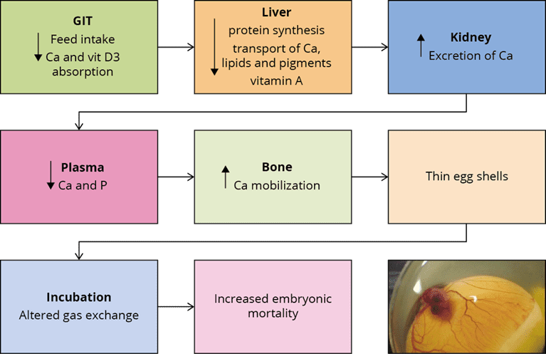
  • Intestine and kidneys: Mycotoxins harm the intestinal epithelium and have nephrotoxic effects, affecting calcium and vitamin D3 absorption and metabolism, necessary for eggshell quality (31). Thin and fragile shells can increase embryonic mortality, lower embryonic weight gain, and hinder hatchability (32).
  • Liver: The liver plays a central role in egg production as it is responsible for vitamin D3 metabolism, the production of nutrient transporters, and the synthesis of the lipids that make up the yolk. Thus, when liver function is impaired, the internal and external quality of the egg declines, which affects DOC production (31-34).
  • Embryo and young chicks: Studies (33-38) have found how mycotoxins affect the embryos. In general, there are two possibilities: the direct one, when the mycotoxin is transferred into the egg, and the indirect one, when the mycotoxin impacts egg quality and, therefore, leads to disease or death of the embryo. The result is a higher embryonic mortality or lower DOC quality. These, among others, result from the lower transfer of antioxidants and antibodies from the hen, low viability of the chick’s immune cells, and higher bacterial contamination. A lower relative weight of the bursa of Fabricio and the thymus is often found.

Qreshi’s team (29) studied the effects on the progeny of broiler breeders consuming feed highly contaminated with AFB1, finding suppression in antibody production and macrophage function in chicks after ten days. Similar results were found by other researchers (36, 37) evaluating the effects of AF and OTA as single and combined contamination. When both mycotoxins are present in the feed, the effect on hatchability and DOC quality are synergistic.

Due to mycotoxin contamination, the reproduction and immune response are impaired, resulting in decreased DOC production and increased early chick mortality, as they are more susceptible to bacterial and viral infections.

Mycotoxins impair table egg production and quality

Studies (22-24) have found mycotoxin contamination in commercial table eggs. A meta-analysis of mycotoxins’ concentration based on 11 published papers was completed recently (22): counting with data from 9509 samples, the meta-analysis reveals an overall presence of mycotoxins in 30% of the samples, being Beauvericin in the first place, followed by DON as well as AF and OTA in third and fourth place, respectively. The risk for humans depends on the intake of contaminated foods in terms of amount and frequency (25), and so far, it has not been estimated in most parts of the world.

Natural contamination in laying hens: a case report

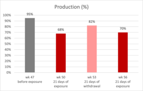
Giancarlo Bozzo’s team (39) reported and published a veterinary case regarding natural mycotoxin contamination in commercial egg production: up to week 47 of age, production parameters were on top of the genetic standards. However, a drop in egg production started at around week 47, and at week 50, egg production was only 68% (figure 1).

Figure
Figure 1: production of laying hens fed naturally contaminated feed with AFB1 and OTA
The house with the reduced performance received feed with linseed. In other houses of the same complex, which did not include linseed in the feed, production was unaffected. Therefore, this raw material was considered a possible cause of the issue. Linseed was removed from the formula, and three weeks after (53 weeks of age), egg production was at 84%. Afterward, linseed got back into the formulation, and the laying rate dropped again to 70% (week 56), this time accompanied by a significant increase in mortality.

Samples were collected at week 56, and AFB1 and OTA were detected in feed and the kidneys and livers of the hens consuming it (table 2). While the levels in the feed were not considered high risk, evidence from necropsy and histopathology suggested either a higher or a prolonged exposure; a synergistic effect of both mycotoxins on hen’s health and productivity can be inferred.

Table 2: mycotoxin analysis results for feed and organs

HPLC analysis results in samples of:
toxin Feed 1
(n=5)
Feed 2
(n=5)
Kidney

(n=10)

Liver

(n=10)

OTA 1.1 ± 0.1 ppb 31 ± 3 ppb 47 ± 3 ppb 24 ± 2 ppb
AFB1 ND 5.6 ± 0.3 ppb 1.4 ± 0.3 ppb 3.6 ± 0.4 ppb

The liver and kidneys were enlarged and showed signs of damage. Furthermore, urate crystals in the peritoneum and the abdominal air sac were observed, indicating renal failure. This limited the excretion of both toxins in the urine, increasing their half-life in the organism and enhancing the effects in target organs, contributing to the synergistic effect observed.

After using mycotoxin-free certified linseed, the problem receded. Though this is the best option to keep animals healthy and productive, it may not be practical in the long term due to the ubiquitous nature of the toxins and the cost and availability constraints of feed raw materials. Moreover, the mycotoxin levels present in the feed were relatively low and fell under recommended guidelines. For these reasons, in-feed toxin mitigation solutions must also be considered to reduce exposure for production animals.

In-feed intervention mitigates the effects of intermittent exposure to multiple mycotoxins

EW Nutrition conducted a study with Hy-Line W-36 layer-breeders intercalating three 10-day cycles of feed with 100ppb AFB1 + 100ppb OTA, with two 21-day cycles of non-challenged feed. An in-feed intervention (Solis Max 2.0, displayed as IFI) containing bentonite, yeast cell wall components, and a mixture of phytogenic components mitigated all effects.

Table 3: experimental groups and mycotoxin challenge

Treatment Group 100 ppb AFB1+ 100 ppb OTA IFI (2 kg/ton)
T-1 Control (C)
T-2 C+IFI X
T-3 Challenge (Ch) X
T-4 Ch+IFI X X

Trial design:

A total of 576 hens (18 replicates per diet, 8 hens each) and 58 roosters were randomly assigned to four diets at 28 weeks of age, as shown in Table 3. The 72-day experimental period included alternating 10-day challenge and 21-day non-challenge intervals (Figure 2). During the challenge intervals, the breeders in T-3 and T-4 were fed the mycotoxin-contaminated feed with and without the IFI.

FigureFigure 2: trial timeline showing challenge and non-challenge intervals and days of data collection and sampling.

Mitigated effects on egg production and egg quality

The challenge decreased overall egg production (Figure 3), egg mass, and shell thickness (Table 4). The first challenge interval did not affect production, but days later, from the first non-challenge period, all parameters were lower for the challenged group.

FigureDifferent letters indicate significant differences (p<0.05). Statistical tendencies (p<0.1) are indicated by (*).

Figure 3: Egg production of hens intermittently challenged with AFB1 and OTA, with and without in-feed Solis Max

The adverse effects on productivity and egg quality started after the first challenged feed was withdrawn and persisted through the following intervals until the end of the experiment. Similar effects in chronic mycotoxin challenges have been previously found (37, 39).

Table 4: Average egg quality parameters of hens intermittently challenged with AFB1+OTA, with and without an in-feed intervention (IFI)

Group Eggshell strength (N) Eggshell thickness (mm) Haugh Units
Control 21,02a 0,3661ab 70,88
IFI 21,16a 0,3702a 71,68
Challenge 20,05b 0,3630b   70,07*
Ch+IFI 21,06a 0,3698a 71,06

Different letters indicate significant differences (p<0.05). Statistical tendencies (p<0.1) are indicated by (*).

Mitigated effects on the progeny in incubation trials

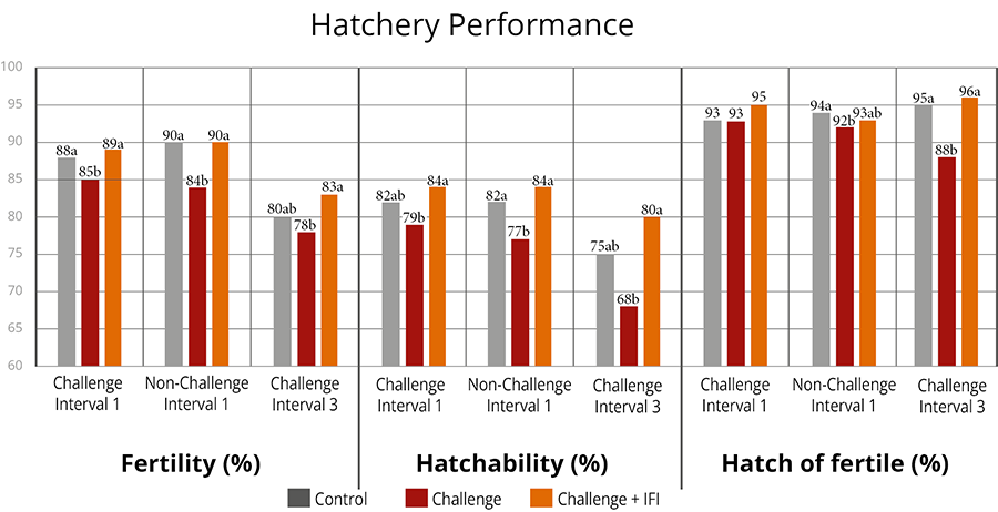
Three incubation trials were performed: after the first challenge and non-challenge interval and at the end of the trial period after the third challenge interval. A significant decrease in fertility and hatchability was observed for the challenged group in all incubation trials. As mycotoxins affect egg quality (22-24) and can be transferred to the eggs (10, 14, 27), the effects were also shown in the case of hatchability and offspring performance. Fertility was affected from the first challenge interval onwards, continuing to be low for the challenge group until the end of the trial. However, the hatchability of fertile eggs dropped after the withdrawal of the contaminated feed and showed the lowest value during the third challenge interval.

The in-feed supplementation of Solis Max 2.0 (IFI) resulted in the consistent recovery of egg production and egg quality throughout the whole experimental period, achieving the same levels of productivity as the non-challenged control.

Figure
Letters indicate significant differences (p<0.05). Statistical tendencies (p<0.1), indicated by (*).

Figure 4: Hatchery parameters of eggs from breeders intermittently challenged with AFB1 and OTA, with and without an in-feed intervention (IFI).

Results in hatch of fertile can be related to egg quality, as the thickness of the eggshell influences the egg’s moisture loss and exchange with the environment during the incubation period. Thinner eggshells lead to higher embryo mortality (31, 32). The group having the challenge with Solis Max showed the same performance as the non-challenged control regarding hatchery performance.

Day-old chick weight was not affected. However, weight gain and mortality after ten days were hindered for the chicks from breeders taking the mycotoxin-contaminated feed (Table 5).

Table 5: Average day- and 10-day-old chick parameters from hens intermittently challenged with AFB1+OTA, with and without an in-feed intervention (IFI)

Parameter Control Challenge Ch + IFI
DOC body weight (g) 36,67 36,24 36,80
10-day body weight (g) 76,30a 75,94b 79,50a
10-day mortality (%) 0,94 1,26 0,97

Letters indicate significant differences (p<0.05). Statistical tendencies (p<0.1) indicated by (*)

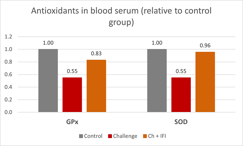
At the end of the experiment, oxidative stress biomarkers were measured in the blood serum of 15 hens per treatment, showing significantly lower GPx, and SOD (figure 5) in the challenged group, which indicates a depletion of the mechanisms to fight oxidative stress (40), the hens taking the in-feed product did not show this depletion.

FigureFigure 5: Antioxidants in blood serum, glutathione peroxidase (GPx), and superoxide dismutase (SOD) from breeders intermittently challenged with AFB1 and OTA, with and without an in-feed intervention (IFI).

Intermittent exposure to AFB1 and OTA negatively affected layer breeder productivity, egg quality, and hatchability and promoted oxidative stress in the birds. Intermittent mycotoxin challenges may affect animals even after the contamination is withdrawn. In-feed interventions showed effectiveness in mitigating these effects.

Climate changes bring new mycotoxin challenges – the right in-feed solutions can help

Today’s mycotoxin scenario shows increased frequency, quantity, and variety. Mainly long-living animals, such as laying hens and breeders, can be at more risk. Additionally, the contamination can be carried over to the eggs, potentially risking human health in the case of table eggs and hatchery performance and DOC quality in the case of breeders.

From case reports, we learn the consequences of real challenges and struggles in commercial production; from scientific trials based on possible commercial situations, we realize the advantages of interventions designed to tackle those challenges.

References

  1. Kos, Jovana, Mislav Anić, Bojana Radić, Manuela Zadravec, Elizabet Janić Hajnal, and Jelka Pleadin. “Climate Change—a Global Threat Resulting in Increasing Mycotoxin Occurrence.” Foods 12, no. 14 (July 14, 2023): 2704. https://doi.org/10.3390/foods12142704.
  2. Zingales, Veronica, Mercedes Taroncher, Piera Anna Martino, María-José Ruiz, and Francesca Caloni. “Climate Change and Effects on Molds and Mycotoxins.” Toxins 14, no. 7 (June 30, 2022): 445. https://doi.org/10.3390/toxins14070445.
  3. Loi, Martina, Antonio F. Logrieco, Tünde Pusztahelyi, Éva Leiter, László Hornok, and István Pócsi. “Advanced Mycotoxin Control and Decontamination Techniques in View of an Increased Aflatoxin Risk in Europe Due to Climate Change.” Frontiers in Microbiology 13 (January 10, 2023). https://doi.org/10.3389/fmicb.2022.1085891.
  4. Medina, Ángel, Jesús M González-Jartín, and María J Sainz. “Impact of Global Warming on Mycotoxins.” Current Opinion in Food Science 18 (December 2017): 76–81. https://doi.org/10.1016/j.cofs.2017.11.009.
  5. Grenier, Bertrand, and Todd Applegate. “Modulation of Intestinal Functions Following Mycotoxin Ingestion: Meta-Analysis of Published Experiments in Animals.” Toxins 5, no. 2 (February 21, 2013): 396–430. https://doi.org/10.3390/toxins5020396.
  6. Filazi, Ayhan, Begum Yurdakok-Dikmen, Ozgur Kuzukiran, and Ufuk Tansel Sireli. “Mycotoxins in Poultry.” Poultry Science, February 15, 2017. https://doi.org/10.5772/66302.
  7. Amirkhizi, Behzad, Seyed Rafie Arefhosseini, Masoud Ansarin, and Mahboob Nemati. “Aflatoxin B1in Eggs and Chicken Livers by Dispersive Liquid–Liquid Microextraction and HPLC.” Food Additives &amp; Contaminants: Part B, August 27, 2015, 1–5. https://doi.org/10.1080/19393210.2015.1067649.
  8. Emmanuel K, Tangni, Van Pamel Els, Huybrechts Bart, Delezie Evelyne, Van Hoeck Els, and Daeseleire Els. “Carry-over of Some Fusarium Mycotoxins in Tissues and Eggs of Chickens Fed Experimentally Mycotoxin-Contaminated Diets.” Food and Chemical Toxicology 145 (November 2020): 111715. https://doi.org/10.1016/j.fct.2020.111715.
  9. Ebrahem, Mohammad, Susanne Kersten, Hana Valenta, Gerhard Breves, and Sven Dänicke. “Residues of Deoxynivalenol (Don) and Its Metabolite de-Epoxy-Don in Eggs, Plasma and Bile of Laying Hens of Different Genetic Backgrounds.” Archives of Animal Nutrition 68, no. 5 (August 20, 2014): 412–22. https://doi.org/10.1080/1745039x.2014.949029.
  10. Salwa, A. Aly, and W. Anwer. “Effect of Naturally Contaminated Feed with Aflatoxins on Performance of Laying Hens and the Carryover of Aflatoxin B1 Residues in Table Eggs.” Pakistan Journal of Nutrition 8, no. 2 (January 15, 2009): 181–86. https://doi.org/10.3923/pjn.2009.181.186.
  11. Devreese, Mathias, Gunther Antonissen, Nathan Broekaert, Siegrid De Baere, Lynn Vanhaecke, Patrick De Backer, and Siska Croubels. “Comparative Toxicokinetics, Absolute Oral Bioavailability, and Biotransformation of Zearalenone in Different Poultry Species.” Journal of Agricultural and Food Chemistry 63, no. 20 (May 19, 2015): 5092–98. https://doi.org/10.1021/acs.jafc.5b01608.
  12. Galtier, P. “Biotransformation and Fate of Mycotoxins.” Toxin Reviews 18, no. 3 (August 1, 1999): 295–312. https://doi.org/10.3109/15569549909162648.
  13. Galtier, P., M. Alvinerie, and J.L. Charpenteau. “The Pharmacokinetic Profiles of Ochratoxin A in Pigs, Rabbits and Chickens.” Food and Cosmetics Toxicology 19 (January 1981): 735–38. https://doi.org/10.1016/0015-6264(81)90528-9.
  14. Hassan, Zahoor Ul, Muhammad Z Khan, Ahrar Khan, Ijaz Javed, and Zahid Hussain. “Effects of Individual and Combined Administration of Ochratoxin A and Aflatoxin B1 in Tissues and Eggs of White Leghorn Breeder Hens.” Journal of the Science of Food and Agriculture 92, no. 7 (December 16, 2011): 1540–44. https://doi.org/10.1002/jsfa.4740.
  15. Li, Shao-Ji, Guangzhi Zhang, Bin Xue, Qiaoling Ding, Lu Han, Jian-chu Huang, Fuhai Wu, Chonggao Li, and Chunmin Yang. “Toxicity and Detoxification of T-2 Toxin in Poultry.” Food and Chemical Toxicology 169 (November 2022): 113392. https://doi.org/10.1016/j.fct.2022.113392.
  16. Prelusky, D.B., R.M.G. Hamilton, and H.L. Trenholm. “Transmission of Residues to Eggs Following Long-Term Administration of 14 C-Labelled Deoxynivalenol to Laying Hens.” Poultry Science 68, no. 6 (June 1989): 744–48. https://doi.org/10.3382/ps.0680744.
  17. Ringot, Diana, Abalo Chango, Yves-Jacques Schneider, and Yvan Larondelle. “Toxicokinetics and Toxicodynamics of Ochratoxin A, an Update.” Chemico-Biological Interactions 159, no. 1 (January 2006): 18–46. https://doi.org/10.1016/j.cbi.2005.10.106.
  18. Osselaere, Ann, Mathias Devreese, Joline Goossens, Virginie Vandenbroucke, Siegrid De Baere, Patrick De Backer, and Siska Croubels. “Toxicokinetic Study and Absolute Oral Bioavailability of Deoxynivalenol, T-2 Toxin and Zearalenone in Broiler Chickens.” Food and Chemical Toxicology 51 (January 2013): 350–55. https://doi.org/10.1016/j.fct.2012.10.006.
  19. Sudhakar, BV. “A Study on Experimentally Induced Aflatoxicosis on the Carryover of Aflatoxin B1 into Eggs and Liver Tissue of White Leghorn Hens.” The Pharma Innovation Journal 11, no. 2S (2022): 213–17.
  20. Yiannikouris, Alexandros, and Jean-Pierre Jouany. “Mycotoxins in Feeds and Their Fate in Animals: A Review.” Animal Research 51, no. 2 (March 2002): 81–99. https://doi.org/10.1051/animres:2002012.
  21. Bouhet, Sandrine, and Isabelle P. Oswald. “The Intestine as a Possible Target for Fumonisin Toxicity.” Molecular Nutrition &amp; Food Research 51, no. 8 (August 2007): 925–31. https://doi.org/10.1002/mnfr.200600266.
  22. Fakhri, Yadolah, Mansour Sarafraz, Amene Nematollahi, Vahid Ranaei, Moussa Soleimani-Ahmadi, Van Nam Thai, and Amin Mousavi Khaneghah. “A Global Systematic Review and Meta-Analysis of Concentration and Prevalence of Mycotoxins in Birds’ Egg.” Environmental Science and Pollution Research 28, no. 42 (September 9, 2021): 59542–50. https://doi.org/10.1007/s11356-021-16136-y.
  23. Osaili, Tareq M., Akram R. Al-Abboodi, Mofleh AL. Awawdeh, and Samah Aref Jbour. “Assessment of Mycotoxins (Deoxynivalenol, Zearalenone, Aflatoxin B1 and Fumonisin B1) in Hen’s Eggs in Jordan.” Heliyon 8, no. 10 (October 2022). https://doi.org/10.1016/j.heliyon.2022.e11017.
  24. Wang, Lan, Qiaoyan Zhang, Zheng Yan, Yanglan Tan, Runyue Zhu, Dianzhen Yu, Hua Yang, and Aibo Wu. “Occurrence and Quantitative Risk Assessment of Twelve Mycotoxins in Eggs and Chicken Tissues in China.” Toxins 10, no. 11 (November 16, 2018): 477. https://doi.org/10.3390/toxins10110477.
  25. Tolosa, J., Y. Rodríguez-Carrasco, M.J. Ruiz, and P. Vila-Donat. “Multi-Mycotoxin Occurrence in Feed, Metabolism and Carry-over to Animal-Derived Food Products: A Review.” Food and Chemical Toxicology 158 (December 2021): 112661. https://doi.org/10.1016/j.fct.2021.112661.
  26. Völkel, Inger, Eva Schröer-Merker, and Claus-Peter Czerny. “The Carry-over of Mycotoxins in Products of Animal Origin with Special Regard to Its Implications for the European Food Safety Legislation.” Food and Nutrition Sciences 02, no. 08 (2011): 852–67. https://doi.org/10.4236/fns.2011.28117.
  27. Yuan, Tao, Junyi Li, Yanan Wang, Meiling Li, Ao Yang, Chenxi Ren, Desheng Qi, and Niya Zhang. “Effects of Zearalenone on Production Performance, Egg Quality, Ovarian Function and Gut Microbiota of Laying Hens.” Toxins 14, no. 10 (September 21, 2022): 653. https://doi.org/10.3390/toxins14100653.
  28. Song, Bin, Teng Ma, Damien P. Prévéraud, Keying Zhang, Jianping Wang, Xuemei Ding, Qiufeng Zeng, et al. “Research Note: Effects of Feeding Corn Naturally Contaminated with Aflatoxin B1, Deoxynivalenol, and Zearalenone on Reproductive Performance of Broiler Breeders and Growth Performance of Their Progeny Chicks.” Poultry Science 102, no. 11 (November 2023): 103024. https://doi.org/10.1016/j.psj.2023.103024.
  29. Qureshi, MA, J Brake, PB Hamilton, WM Hagler, and S Nesheim. “Dietary Exposure of Broiler Breeders to Aflatoxin Results in Immune Dysfunction in Progeny Chicks.” Poultry Science 77, no. 6 (June 1998): 812–19. https://doi.org/10.1093/ps/77.6.812.
  30. Ul-Hassan, Zahoor, Muhammad Zargham Khan, Ahrar Khan, and Ijaz Javed. “Immunological Status of the Progeny of Breeder Hens Kept on Ochratoxin a (OTA)- and Aflatoxin B1(Afb1)-Contaminated Feeds.” Journal of Immunotoxicology 9, no. 4 (April 24, 2012): 381–91. https://doi.org/10.3109/1547691x.2012.675365.
  31. Devegowda, G., and D. Ravikiran. “Mycotoxins and Eggshell Quality: Cracking the Problem.” World Mycotoxin Journal 1, no. 2 (May 1, 2008): 203–8. https://doi.org/10.3920/wmj2008.1037.
  32. Onagbesan, O., V. Bruggeman, L. De Smit, M. Debonne, A. Witters, K. Tona, N. Everaert, and E. Decuypere. “Gas Exchange during Storage and Incubation of Avian Eggs: Effects on Embryogenesis, Hatchability, Chick Quality and Post-Hatch Growth.” World’s Poultry Science Journal 63, no. 4 (December 1, 2007): 557–73. https://doi.org/10.1017/s0043933907001614.
  33. Ebrahem, Mohammad, Susanne Kersten, Hana Valenta, Gerhard Breves, Andreas Beineke, Kathrin Hermeyer, and Sven Dänicke. “Effects of Feeding Deoxynivalenol (Don)-Contaminated Wheat to Laying Hens and Roosters of Different Genetic Background on the Reproductive Performance and Health of the Newly Hatched Chicks.” Mycotoxin Research 30, no. 3 (April 11, 2014): 131–40. https://doi.org/10.1007/s12550-014-0197-z.
  34. Yegani, M., T.K. Smith, S. Leeson, and H.J. Boermans. “Effects of Feeding Grains Naturally Contaminated with Fusarium Mycotoxins on Performance and Metabolism of Broiler Breeders.” Poultry Science 85, no. 9 (September 2006): 1541–49. https://doi.org/10.1093/ps/85.9.1541.
  35. Calini, F, and F Sirri. “Breeder Nutrition and Offspring Performance.” Revista Brasileira de Ciência Avícola 9, no. 2 (June 2007): 77–83. https://doi.org/10.1590/s1516-635×2007000200001.
  36. Hassan, ZU, MZ Khan, A Khan, I Javed, U Sadique, and A Khatoon. “Ochratoxicosis in White Leghorn Breeder Hens: Production and Breeding Performance.” Vet. J. 32, no. 4 (2012): 557–61.
  37. Verma, J., T. S. Johri, and B. K. Swain. “Effect of Varying Levels of Aflatoxin, Ochratoxin and Their Combinations on the Performance and Egg Quality Characteristics in Laying Hens.” Asian-Australasian Journal of Animal Sciences 16, no. 7 (January 1, 2003): 1015–19. https://doi.org/10.5713/ajas.2003.1015.
  38. Johnson-Dahl, M.L., M.J. Zuidhof, and D.R. Korver. “The Effect of Maternal Canthaxanthin Supplementation and Hen Age on Breeder Performance, Early Chick Traits, and Indices of Innate Immune Function.” Poultry Science 96, no. 3 (March 2017): 634–46. https://doi.org/10.3382/ps/pew293.
  39. Bozzo, Giancarlo, Nicola Pugliese, Rossella Samarelli, Antonella Schiavone, Michela Maria Dimuccio, Elena Circella, Elisabetta Bonerba, Edmondo Ceci, and Antonio Camarda. “Ochratoxin A and Aflatoxin B1 Detection in Laying Hens for Omega 3-Enriched Eggs Production.” Agriculture 13, no. 1 (January 5, 2023): 138. https://doi.org/10.3390/agriculture13010138.
  40. Surai, Peter F., Ivan I. Kochish, Vladimir I. Fisinin, and Michael T. Kidd. “Antioxidant Defence Systems and Oxidative Stress in Poultry Biology: An Update.” Antioxidants 8, no. 7 (July 22, 2019): 235. https://doi.org/10.3390/antiox8070235.


Effects on Performance and Gut Health of Ventar D Supplementation in Broiler Diets

Summary of study by Necmettin Ceylan, Sait Koca, Nejla Kahraman, Ankara University, Faculty of Agriculture, Animal Science, 6110 Ankara/Türkiye

The study conducted by Dr. Celyn et al. in 2023 focused on the impact of Ventar D supplementation in broiler diets on growth performance and gut health. The trial was carried out over six weeks on Ross 308 broiler chicks, comparing a control group with an experimental group supplemented with Ventar D. The trial feed was based on corn, soybean meal, wheat, sunflower meal, and poultry oil.

Key Findings

Growth Performance: The study demonstrated that Ventar D supplementation significantly improved body weight gain, feed consumption, feed conversion ratio (FCR) and EPEF during the starter, grower, and finisher periods. The overall performance of chickens fed with Ventar D was notably better, showing a 6.5% higher body weight and 1.67% better FCR compared to the control group.

Treatments BWG, g FCR Corrected FCR2565 FI, g Mortality,% EPEF
Control 2520.6a±32,77 1.620a±0.006 1.629a±0.011 4082.2a±46.77 3.25±0.28 367.2a±5.18
Ventar D 2684.3b±23.65 1.593b±0.010 1.568b±0.015 4273.9b±19.89 2.75±0.53 399.8b±4.35

Different letters indicate significance; P ≤ 0.05

Liver Enzymes: The addition of Ventar D led to a significant decrease in serum Alanine aminotransferase (ALT) levels

Treatments ALP ALT
Control 286.70±54.98 1.505a±0.390
Ventar D 301.50±87.19 0.832b±0.181

Different letters indicate significance; P ≤ 0.05

Gut Health: Ventar D supplementation resulted in higher concentrations of short-chain volatile fatty acids (SCVFA) in the cecum.

  Acetate Propionate Butyrate Isobutyrate Valerate Isovalerate BCFA Total SCFA
Control 27.22a±1.26 8.21±0.38 7.24a±0.41 0.848±0.078 0.964±0.043 0.881±0.054 2.69a±0.12 45.36a±1.53
Ventar D 30.51b±0.80 9.36±0.56 8.86b±0.44 0.878±0.070 1.121±0.077 0.993±0.031 2.99b±0.08 51.73b±1.32

Different letters indicate significance; P ≤ 0.05

Conclusion

Considering the results summarized in the tables above according to the feeding phases and the overall study (0-41 days): Ventar D supplementation of broiler feeds at the level of 100 g/ton significantly improved growth performance parameters during the starter, grower and finisher periods (P ≤ 0.05), and in the final results was stable at 6.5% higher BW and 1.67% better FCR compared to the control group. European Production Efficiency Factor (EPEF) was also significantly better than the control group (P ≤ 0.05).

In the study, liver enzyme and the concentration of short-chain volatile fatty acids also improved significantly with the addition of Ventar D, which may be attributed to the gut health related mode of action for Ventar D.


Low Crude Protein Diets in Poultry: Understanding the Consequences

BROILER

Conference report

The concept of feeding poultry, specifically broilers and layers, with reduced crude protein (CP) diets is gaining traction among nutritionists. The economic implications of balancing amino acids currently dictate dietary CP levels. At the recent EW Nutrition Poultry Academy in Jakarta, Indonesia, Dr. Steve Leeson, Professor Emeritus at the University of Guelph, Canada, raised a crucial question: “What does ‘low CP’ really mean?” He states that it typically means a reduction of maximum 2-3% relative to current CP levels.

Low CP diets generally involve a decrease in soybean meal, compensated by higher grain content. This change increases dietary starch and decreases dietary lipid levels. To meet nutritional needs, these diets also include higher amounts of crystalline (synthetic) amino acids.

Dr. Leeson outlined the advantages and disadvantages of low CP diets. Positives include improved gut health due to reduced proteolytic bacteria, less environmental pollution, lower water intake (improving litter quality), improved sustainability indices, increased dietary net energy, and better performance during heat stress. Negatives encompass issues like lower pellet quality, altered dietary electrolyte balance, higher diet costs, reduced growth rate and feed efficiency, and increased abdominal fat deposition. There are also questions about the presumed complete utilization of crystalline amino acids, which can be as high as 25kg/MT in these diets.

Challenges with Low CP Diets

  • Protein vs. Amino Acids: Diets are typically formulated based on digestible amino acid content, though minimum CP levels remain common, to avoid reduced performance: Dr. Leeson noted that broiler diets with less than 19% CP in starter and 15% in finisher phases, and layer diets below 13% CP, often fail to deliver adequate performance, regardless of digestibly amino acid supply.
  • Utilization of Free Amino Acids: The crystalline amino acids are immediately absorbable in the small intestine, contrasting with protein-bound amino acids that are absorbed as di- and tri-peptides. Amino acids absorption dynamics and endogenous loss of amino acids are affected by (high) levels of  crystalline amino acids.
  • Non-Essential Amino Acids: The impact of reduced CP on animal performance might be related to the lower levels of presumed non-essential amino acids, e.g. glycine and serine.  This is an area for further exploration.
  • Energy Level Considerations: Dr. Leeson suggests maintaining specific ratios of digestible lysine to apparent metabolizable energy in broilers at different growth stages. The heat increment of CP is an essential factor, as it reduces net energy efficiency, possibly requiring an adjustment in amino acid to metabolizable energy ratios as poultry diets are not based on net energy values.
  • Gut Health: Lower CP levels can reduce the flow of undigested protein into the hindgut, reducing the risk of necrotic enteritis, and the production of harmful metabolites, like biogenic amines.
  • Role of Proteases: Protease use can lead to a further 2-4% reduction in dietary CP, with the response depending on the inherent protein digestibility of the diets.
  • Impacts on Pellet Quality: Due to the binding properties of protein, each 1% reduction in CP typically results in a 2% decrease in pellet durability (index).
  • Electrolyte Balance: Reduced CP can significantly lower dietary electrolyte balance, which has to be considered in feed formulation. Amongst the nutrients contributing to DEB value, Sodium and Potassium appear to be the most influential minerals to consider.

Conclusion

Dr. Leeson anticipates that low CP diets will become increasingly relevant. They have the potential to reduce environmental pollution and dependence on soybean meal, despite current challenges in reducing feed costs.


***

EW Nutrition’s Poultry Academy, featuring Dr. Leeson, took place in Jakarta and Manila in early September 2023. With nearly 50 years of industry experience, Dr. Leeson has made significant contributions to poultry nutrition and management, evidenced by his numerous awards and over 400 published papers.


Nutritional considerations for immunity and gut health

Chicks
Conference report 

At the recent EW Nutrition Poultry Academy in Jakarta, Indonesia, Dr Steve Leeson, Professor Emeritus, University of Guelph, Canada, opened his presentation by stating that “it is obvious that any nutrient deficiency will impact bird health, but not so obvious is that nutrition per se can positively impact immunity and health in an otherwise healthy and high-producing bird.”

Modern high-performing broilers are characterized by extremely high feed intake. This puts a lot of stress on the physiology of the entire gastrointestinal tract, but particularly so on the absorptive epithelial cells of the small intestine. Any organism requires a nutrient source for survival and reproduction. Dr Leeson asked “can we significantly reduce nutrient supply to pathogens, while sustaining bird productivity?”

He reminded the audience that no cellular function comes for free: so there is always a “cost”. A general conclusion is that 10% of nutrients can be used for immune function during disease challenge, and always get priority. Therefore, you don’t want to overstimulate the immune system, which in extreme situations leads to an inflammatory response. In his presentation, Dr Leeson considered factors determining gut health and nutritional tools which are available to support gut health.

Gut microflora

Gut pathogens impact the bird and/or the consumer. Clostridia and E. coli are the major concerns regarding bird health and productivity, whereas Salmonella and Campylobacter are major pathogens important for human health.

The chick hatches with a gut virtually devoid of microbes, so early colonizers tend to predominate quite quickly. Microbial species present on the hatching tray, during delivery and during the first few days at the farm will likely dictate early gut colonization. In some instances, the chick’s microflora may be established by the time it gets to the farm, so the probiotic faces more of a challenge to establish itself as the predominant species.

Antibiotic alternatives

Gut villi development matures at around 10-15 days of age. The broiler pre-starter diet therefore is a target for feed additives that positively impact gut structure and development.

  • Among the short chain fatty acids, butyric acid is considered the prime energy source for enterocytes and it is also necessary for the correct development of the gut-associated lymphoid tissue (GALT). Butyric acid can also be added indirectly via fermentation of judicious levels of soluble fiber to encourage optimal gut villi development. Dr Leeson added that he is a big believer in butyric acid, encouraging a good gut structure at 10 days, which can be worth about 50 kcal.
  • Exogenous enzymes should also be considered in an attempt to maximize digestion and limit the flow of nutrients to the large intestine and ceca. Protease enzymes have great potential in this regard, since they allow nutritionists to reduce dietary crude protein and hopefully reduce the supply of nitrogen that fuels proteolytic Clostridia bacteria in the large intestine and ceca.
  • Amino acids, particularly threonine, play a critical role in the maintenance of intestinal mucosal integrity and barrier function, especially for mucin synthesis, which protects enterocytes from adherence by pathogenic bacteria, and from attack by endogenous enzymes and acids.
  • Polyunsaturated fatty acids (PUFAs) – Omega-3s and especially DHA from fish oil help to reduce inflammatory response (overstimulation). Omega-3s are poorly converted to DHA by the chicken, so conventional sources such as flax are of limited application for immunity.
  • Blood plasma from pigs or cattle is a complex spray-dried mixture of proteins and amino acids, many of which are immunoglobulins that “temper” the immune system, much like PUFAs.
  • Vitamins A, D, E and C have vital roles in the normal function of the immune system and have antioxidant capacity.
  • Certain complex carbohydrates, such as ß-glucans, influence gut health due to their fermentation, leading to the production of short-chain fatty acids, such as butyrate.
  • Antioxidants – to firstly control oxidation of fats and fat-soluble vitamins in feed, and secondly to optimize birds’ cellular oxidative capacity, to prevent cell damage, therefore maintaining healthy cellular and immune function.
  • Betaine increases intracellular water retention, reducing “dehydration” of microvilli and increasing their volume/surface area.
  • Fiber – moderate levels (1-2%) of soluble (fermentable) and insoluble fiber can be beneficial to early gut development by stimulating gizzard development and endogenous enzyme production.
  • Phytogenics are becoming very common in combination with acidifiers (upper tract) and probiotics. Essential oils are becoming more mainstream the more we know about them.

Recommendations for optimizing gut health and immunity

Fast growth rate and high egg output are negatively correlated with immune response. Consequently, nutrient-dense diets are not optimal for immunity. With bacteria, it’s a numbers game – but these numbers quickly multiply. The first 7 days are important, therefore probiotics must be established early. Consider the role of targeted feed additives, such as butyrate, phytogenics, antioxidants, PUFAs etc.

Also, maximize feed particle size – the limit is usually pellet quality. Mitigate nutrient transition at any diet change. Review the supply of trace minerals, as there is a trend to lower levels of organic minerals. With all the factors that weigh into production performance, any support that can be rallied through nutrition needs to be considered.


***

EW Nutrition’s Poultry Academy took place in Jakarta and Manila in early September 2023. Dr. Steve Leeson, an expert in Poultry Nutrition & Production with nearly 50 years’ experience in the industry, was the distinguished keynote speaker.

Dr. Leeson had his Ph.D. in Poultry Nutrition in 1974 from the University of Nottingham. Over a span of 38 years, he was a Professor in the Department of Animal &Poultry Science at the University of Guelph, Canada. Since 2014, he has been Professor Emeritus at the same University. As an eminent author, he has more than 400 papers in refereed journals and 6 books on various aspects of Poultry Nutrition & Management. He also won the American Feed Manufacturer’s Association Nutrition Research Award (1981), the Canadian Society of Animal Science Fellowship Award (2001), and Novus Lifetime Achievement Award in Poultry Nutrition (2011).


Decoding the connection between stress, endotoxins, and poultry health

Poultry

By Marisabel Caballero, Global Technical Manager Poultry, EW Nutrition

Stress can be defined as any factor causing disruptions to homeostasis, which triggers a biological response to regain equilibrium. We can distinguish four major types of stressors in the poultry industry:

  • Technological: related with management events and conditions
  • Nutritional: involving nutritional disbalances, feed quality and feed management
  • Pathogenic: comprising health challenges.
  • Environmental: changes in environment conditions

In practical poultry production, multiple stress factors occur simultaneously. Their effects are also additive, leading to chronic stress. The animals are not regaining homeostasis and continuously deviate the use of resources through inflammation and the gut barrier-function, thus leading to microbiome alteration. As a consequence, welfare, health, and productivity are compromised.

What are endotoxins?

Bacterial lipopolysaccharides (LPS), also known as endotoxins, are the main components of the outer membrane of all Gram-negative bacteria and are essential for their survival. LPS have direct contact with the bacteria’s surroundings and function as a protection mechanism against the host’s immunological response and chemical attacks from bile salts, lysozymes, or other antimicrobial agents.

Gram-negative bacteria are part of animals’ microbiota; thus, there are always LPS in the intestine. Under optimal conditions, this does not affect the animals, because intestinal epithelial cells are not responsive to LPS when stimulated from the apical side. In stress situations, the intestinal barrier function is impaired, allowing the passage of endotoxins into the blood stream. When LPS are detected by the immune system either in the blood or in the basolateral side of the intestine, inflammation and changes in the gut epithelial structure and functionality occur.

The gut is critically affected by stress

Even when there is no direct injury to the gut, signals from the brain can modify different functions of the intestinal tract, including immunity. Stress can lead to functional disorders, as well as to inflammation and infections of the intestinal tract. Downstream signals act via the brain–gut axis, trigger the formation of reactive oxygen and nitrogen species as well as local inflammatory factors, and circulating cytokines, affecting intestinal homeostasis, microbiome, and barrier integrity.

Stress then results in cell injury, apoptosis, and compromised tight junctions. For this reason, luminal substances, including toxins and pathogens, leak into the bloodstream. Additionally, under stress, the gut microbiome shows and increment on Gram-negative bacteria (GNB). For instance, a study by Minghui Wang and collaborators (2020) found an increase of 24% in GNB and lower richness, in the cecum of pullets subjected to mild heat stress (increase in ambient temperature from 24 to 30°C).

Both these factors, barrier damage and alterations in the microbiome, facilitate the passage of endotoxins into the blood stream, which promotes systemic chronic inflammation.

What categories of stress factors trigger luminal endotoxins’ passage into the bloodstream?

Technological stress

Various management practices and events can be taken as stressors by the animals’ organism. One of the most common examples is stocking density, defined as the number of birds or the total live weight of birds in a fixed space. High levels are associated with stress and loss of performance.

A study from the Chung-Ang University in 2019 found that broilers with a stocking density of 30 birds/m2 presented two times more blood LPS than birds kept at half of this stocking density. Moreover, the body weight of the birds in the high-density group was 200g lower than the birds of the low-density group. The study concluded that high stocking density is a factor that can disrupt the intestinal barrier.

Nutritional stress

The feed supplied to production animals is designed to contribute to express their genetic potential, though some feed components are also continuous inflammatory triggers. Anti-nutritional factors, oxidized lipids, and mycotoxins induce a low-grade inflammatory response.

For instance, when mycotoxins are ingested and absorbed, they trigger stress and impair immunity in animals. Their effects start in gastrointestinal tract and extend from disrupting immunity to impairing the intestinal barrier function, prompting secondary infections. Mycotoxins can increase the risk of endotoxins in several ways:

A study conducted by EW Nutrition (Figure 1) shows an increase in intestinal lesions and blood endotoxins after a mycotoxin challenge of 200pbb of Aflatoxin B1 + 360ppb Ochratoxin in broilers at 21 days of age. The challenged birds show two times more lesions and blood endotoxins than the ones in the unchallenged control. The use of the right mitigation strategy, a product based on bentonite, yeast cell walls, and phytogenics (EW Nutrition GmbH) successfully prevented these effects as it not only mitigates mycotoxins, but also targets endotoxins in the gut.

Figure 1
Figure 1 Blood LPS and intestinal lesion score of broilers challenged with 200ppb AFB1 + 350 ppb OTA from 1 to 21 days of age without and with an anti-toxin product from EW Nutrition GmbH (adapted from Caballero et al., 2021)

Pathogenic stress

Intestinal disease induces changes in the microbiome, reducing diversity and allowing pathogens to thrive. In clinical and subclinical necrotic enteritis (NE), the intestinal populations of GNB, including Salmonella and E.coli also increases. The lesions associated with the pathogen compromise the epithelial permeability and the intestinal barrier function, resulting in translocation of bacteria and LPS (Figure 5) into the bloodstream and internal organs.

Figure 2
Figure 2 Increase in systemic LPS (vs a healthy control) after a NE challenge (adapted from Chen et al., 2015, Li et al., 2018 & Liu at al., 2018)

Environmental stress

Acute and chronic heat and cold stress increases gut permeability, by increasing intestinal oxidative stress and disrupting the expression of tight junction proteins. This results in the damage and destruction of intestinal cells, inflammation, and imbalance of the microbiota. An increased release and passage of endotoxins has been demonstrated in heat stress (Figure 3), as well as a higher expression of TLR-4 and inflammation.

Figure 3
Figure 3 Systemic LPS increase (in comparison with a non-stressed control) after different heat stress challenges in broilers: 16°C increased for 2, 5 and 10 hours (Huang et al., 2018); 9°C increased for 24 and 72 hours (Nanto-Hara et al., 2020); 10°C continuously for 3 and 10 days, and 15°C 4 hours daily for 3 and 10 days (Alhenaky et al., 2017)


Zhou and collaborators (2021) showed that 72 hours of low temperature treatment in young broilers increased intestinal inflammation and expression of tight junction proteins, while higher blood endotoxins indicate a disruption of the intestinal barrier. As a consequence, the stress decreased body gain and increased the feed conversion rate.

An experiment conducted by EW Nutrition GmbH with the objective of evaluating the ability of a toxin mitigation product to ameliorate heat-stress induced LPS. For the experiment, 1760 Cobb 500 pullets were divided into two groups, and each was placed in 11 pens of 80 hens, in a single house. One of the groups received feed containing 2kg/ton of the product from the first day. From week 8 to week 12, the temperature of the house was raised 10°C for 8 hours every day.

Throughout the heat stress period, blood LPS (Fig 4) was lower in the pullets receiving the product, which allowed lower inflammation, as evidenced by the lower expression of TLR4 (Fig. 5). Oxidative stress was also mitigated with the help of the combination of phytomolecules in the product, obtaining 8.5% improvement on serum total antioxidant capacity (TAC), supported by an increase in in superoxide dismutase (SOD glutathione peroxidase (GSH) and a decrease in malondialdehyde (MDH).

Fig

Figures 4-5
Figures 4 and 5 – Blood LPS and expression of toll-like receptor 4 (TLR4) in lymphocites of pullets before (wk 6) and during heat stress (wk 9 and 10). (*) indicates significant differences (P<0,05), and (‡) a tendency to be different against the control group (P<0,1).

In practice: there is no silver bullet

In commercial poultry production, a myriad stressors may occur at the same time and some factors trigger a chain of events that work to the detriment of animal health and productivity. Reducing the solution to the mitigation of LPS is a deceitfully simplistic approach. However, this should be part of a strategy to achieve better animal health and performance. In fact, EW Nutrition’s toxin mitigation product alone helped the pullets to achieve 3% improvement in body weight and 9 points lower cumulative feed conversion (Figure 6).

Figure 6
Figure 6

Keeping the animals as free of stress as possible is a true priority for poultry producers, as it promotes animal health as well as the integrity and function of the intestinal barrier. Biosecurity, good environment, nutrition and good management practices are crucial; the use of feed additives to reduce the consequences of unavoidable stress also critically supports the profitability of poultry operations.


Coccidiosis management without increasing antimicrobial resistance – it’s up to us

Header POULTRY BROILERS Broilers

By Tingting Fan, Regional Technical Manager Poultry, EW Nutrition

Chicken coccidiosis is a common and important disease in poultry production, with an incidence of infection as high as 50-70%. The mortality rates are around 20-30% or higher in highly severe cases. In addition to losses due to mortality, producers lose money due to poor growth as well as decreased meat yield and quality. Additionally, the birds get more susceptible to secondary infections, e.g., necrotic enteritis (Moore, 2016).

The costs caused by coccidiosis in poultry are about 13 billion US $ (Blake, 2020). These costs globally divide into 1 billion costs for prophylaxis/treatment and 12 billion due to performance losses. Until now, only 5% of the prophylaxis costs have been created by natural solutions. That means that there is still a high potential to be tapped.

Natural solutions, unfortunately, are only used by a minority

For a long time, ionophores fitting the classical definition of antibiotics and chemicals were used in coccidia-fighting programs – and contributed to the development of antimicrobial resistance (Nesse et al., 2015). Nowadays, the combination with vaccination in rotation or shuttle programs has reduced this danger, but there is still potential. Meanwhile, some natural solutions are available that can be integrated into coccidiosis-fighting programs. However, producers using natural solutions are still a minority.

For thousands of years, plants have been used in human and veterinary medicine. Before the discovery of antibiotics in 1928, diseases were fought with plants. To regain the effectiveness of antibiotics, using natural solutions for prophylaxis should be once more standard, and the use of antibiotics is the treatment only for critical cases.

How does Eimeria damage broilers

The pathogenic mechanism of coccidia or Eimeria spp. is mainly the massive destruction of host intestinal cells when it reproduces, resulting in severe damage to the intestinal mucosa. On the one hand, the damaged gut wall loses its capability for effective digestion and absorption of nutrients, leading to worse feed conversion and lower weight gain.

On the other hand, this damage reduces the chicken’s immunity and paves the way for other infections, such as necrotic enteritis, and raises mortality.

Table 1:The seven most known Eimeria species in broilers and their main site of occurrence

Eimeria species Predilection site
E. tenella Ceca
E. acervulina Duodenum and prox. jejunum
E. maxima Central jejunum
E. mitis Distal jejunum and ileum
E. necatrix Central jejunum and ceca
E. brunetti Ileum, entrance of the ceca and rectum
E. praecox Duodenum and prox. jejunum

Concerning their pathogenicity, for poultry, the Eimeria species must be ordered in the following way: E. necatrix> E. tenella > E. brunetti > E. maxima > E. acervulina > Eimeria mitis, and Eimeria praecox.

Prevention is better than treatment

Thanks to its bi-layered wall with a robust structure, the oocysts of coccidia are extremely resilient. They can survive 4 to 9 months in the litter or soil and are resistant to common disinfectants. Farm personnel and visitors are also important vectors, so good biosecurity practices can reduce the number of oocysts contaminating the premises and help prevent clinical out-brakes. Coccidiosis control in poultry should focus on “prevention” rather than “treatment”, combining biosecurity practices, feed additives, and/or vaccination.

Effective hygiene on the farm is crucial

To prevent coccidia infections, one of the most critical points is hygiene. Biosecurity practices are crucial and include cleaning and disinfection of the poultry houses and their surroundings, pest control and prevention, restriction, control, and management of the entry of personnel, visitors, vehicles, and equipment, among others.

Coccidia oocysts are ubiquitous and survive for a long time, and even effective cleaning and disinfection cannot completely remove them. After a severe outbreak, it is recommended to take drastic biosecurity measures such as flame or caustic soda disinfection to prevent further spread of the disease.

When there are birds in the house, it must be paid attention that the litter is not excessively humid. Litter moisture should be maintained around 25%; turning and replacing moist litter are the best practices to follow. For keeping the litter dry, adequate ventilation and appropriate stocking density are beneficial.

To avoid unnecessary stress and gut health issues, the birds must be fed according to their requirements with high-quality feed so that the animals build up good immunity and resilience.

Coccidiosis can be controlled with effective programs

Anticoccidial drugs were the first means of preventing and controlling coccidiosis in chickens and once achieved very good results. Since Sulfaquinoxaline was found to be effective in the 1850s, about fifty other drugs have been developed for the prevention and control of coccidiosis. Generally, the anticoccidials used for years to prevent the disease can be divided into ionophores and chemicals.

Ionophores, produced as by-products of bacterial fermentation, are technically antibiotics. The great benefits of ionophores are that they kill the parasite before it can infect the bird and thus prevent damage to the host cells. Eimeria species also take a long time to develop resistance to ionophores (Chapman, 2015). Well-established ionophores are products that contain monensin, lasalocid, salinomycin, narasin, or maduramycin; the trade names are Coban/Monensin, Avatec, Coxisstac, Monteban, and Cygro.

Chemicals, these molecules, are produced by chemical synthesis. They differ from each other and ionophores as each one has a unique mode of action against coccidia. In general, they act by interfering with one or more stages of the life cycle of Eimeria, e.g., supplying fake nutrients (Amprolium, Vit. B1) to the parasite, starving them out. The active components here are nicarbazin, amprolium, zoalene, decoquinate, clopidol, robenidine and diclazuril, and the respective trade names Nicarb, Amprol, Zoamix, Deccox, Coyden, Robenz and Clinacox. Eimeria species develop resistance to these chemical molecules; therefore, they must be used carefully and with strict planning. However, cross-resistance does not develop, making them highly valuable in rotation programs.

Vaccination against coccidiosis is accepted by many farmers as a good solution to control coccidiosis in chickens. Vaccination aims to replace resistant field strains with vaccine strains, which are sensitive to anticoccidials. Currently, commercial chicken vaccines are available in natural and attenuated strains; research to obtain safer and more efficient vaccines is also ongoing.

Non-attenuated vaccines are less expensive and make for good immunity, but as they may mildly damage the intestinal epithelium, the risk of necrotic enteritis can increase. On the contrary, attenuated strains – usually “precocious” strains with shorter reproduction cycles, cause less intestinal damage and thus have a lower risk of provoking bacterial or necrotic enteritis. The immunity is like after normal infections; however, you have a controlled epidemiology, fewer coccidiosis outbreaks, and an improved uniformity of the flock.

Phytomolecules-based natural anticoccidials saponins and tannins are natural components that can also help control coccidiosis (e.g., Pretect D, EW Nutrition GmbH). These ingredients act in different ways: the tannins improve the intestinal barrier function locally and systemically. The saponins directly impact the oocysts by preventing their growth, interacting with the cholesterol in the cell membrane (triterpenoid saponin), or hindering further sporulation and causing cell death by causing pores in the cell membrane of the parasite. Altogether, Pretect D promotes the beneficial microbial population and reduces the harmful one, improves the gut barrier function, reduces mucosal inflammation, inhibits growth and replication of Eimeria, preventing their lesions, and fosters birds’ immune response against Eimeria spp.

To prove Pretect D’s effectiveness in the reduction of coccidiosis, several trials were conducted. One of the trials was carried out in Poland with 360.000 broilers in commercial conditions. The animals were divided into ten houses, and two cycles were tested. Half of the birds served as control and received Narasin and Nicarbazin in the starter and grower I diet and salinomycin in the grower II diet. The other half also were fed Narasin and Nicarbazin in the starter and grower I diet, but Pretect D @1kg/t in grower II and 0.5kg/t in the finisher diet. The results are shown in figure 1: The application of Pretect D in the grower II and finisher diet decreased the number of oocysts in the droppings more than the application of salinomycin and, therefore, reduced the spreading of coccidiosis. In addition, the performance of the broilers receiving Pretect D was nothing short of the control’s performance showing Pretect as an optimal completion in shuttle or rotation programs (see more HERE).

Figure Reduction Of Oocysts In The Droppings By Pretect DFigure 1: Reduction of oocysts in the droppings by Pretect D

Managing coccidiosis without promoting antimicrobial resistance is not easy, but feasible

Coccidiosis is a challenge aggravated by our current high level of production. Tools such as ionophores, chemicals, but also vaccines, and natural products are available to fight coccidiosis. However, due to the high probability of resistance development, these tools must be used carefully and in structured programs. The phytomolecules-based product Pretect D gives the possibility to reduce antimicrobial resistance as part of programs against coccidiosis.

References upon request


Pathogenic Enterococcus cecorum – an emerging profit killer for broiler producers

Header

By Dr. Ajay Bhoyar, Global Technical Manager, EW Nutrition

Pathogenic Enterococcus cecorum (EC) is emerging as a significant challenge in poultry production worldwide, causing substantial losses to commercial flocks. EC has become a considerable concern for the poultry industry, not only because of its rapid spread and negative impact on broiler health but also because of its increasing antibiotic resistance. As a result, there is a growing need to explore alternative ways of controlling this bacterium. There is no silver bullet yet as a replacement for antibiotics to limit the load of E. cecorum. Maintaining optimum gut health to avoid E. cecorum leakage during the first week of the broiler’s life can control losses due to E. cecorum.

Phytogenic compounds, which are derived from plants, have gained attention in the last decades as a potential solution for controlling common gut pathogens. These natural compounds have been found to possess antimicrobial properties and can help improve gut health in broilers. In this article, we will discuss the current state of E. cecorum and explore potential strategies, including using phytogenic compounds as support in controlling economic losses due to this emerging pathogen in broiler production.

Enterococcus cecorum and its negative impact on broiler production

E. cecorum is a component of normal enterococcal microbiota in the gastrointestinal tract of poultry. These are facultatively anaerobic, gram-positive cocci. Over the past 15 years, pathogenic strains of E. cecorum have emerged as an important cause of skeletal disease in broiler and broiler breeder chickens (Broast et al., 2017; Jackson et al., 2004 and Jung et al., 2017). Along with the commensal strains of EC, the pathogenic strains also occur and can result in Enterococcal spondylitis (ES), also known as “kinky back”, a serious disease of commercial poultry production in which the bacteria translocate from the intestine to the free thoracic vertebrae and adjacent notarium or synsacrum, causing lameness, hind-limb paresis and, in 5 to 15% of cases, mortality (de Herdt et al., 2008; Martin et al., 2011; Jung and Rautenschlein, 2014). The compression of the spinal cord due to infection of the free thoracic vertebra results in the so-called “kinky back” in the skeletal phase of E. cecorum infection. Kinky back is also a common name for spondylolisthesis, a developmental spinal anomaly. EC is normally found in the gastrointestinal tract and may need the help of other factors, such as a leaky gut, to escape the gastrointestinal tract. The emergent pathogenic strains of E. cecorum have developed an array of virulence factors that allow these strains to 1) colonize the gut of birds in the early life period; 2) escape the gut niche; 3) spread systemically while evading the immune system; and 4) colonize the damaged cartilage of the free thoracic vertebra (Borst, 2023). The E. cecorum can invade internal organs and produce lesions in the pericardium, lung, liver, and spleen.

The negative impact of E. cecorum on broiler economics, health, and welfare

Enterococcus cecorum can harm broiler health, welfare, and economics. This can result in decreased profitability for broiler producers.

The broiler flocks infected with E. cecorum may have reduced feed intake/ nutrient absorption and reduced growth rates, leading to a higher feed conversion ratio, longer production cycles, and lower weight gain. The morbidity and mortality from E. cecorum infection can be as high as 35 % and 15%, respectively. The higher condemnations of up to 9.75% at the processing plant can further add to the losses (Jung et al., 2018).  This can result in significant economic losses for producers.

Further, E. cecorum infections can impair the immune function of broilers, making them more susceptible to other pathogens and reducing their overall health and welfare. Pathogenic E. cecorum is an opportunistic pathogen that can gain momentum during coinfection with E. coli and other gut pathogens, causing a leaky gut. Therefore, a holistic approach to gut health management may help reduce the losses.

E Cecorum

Antibiotic resistance in E. cecorum

E. cecorum has been found to be resistant to multiple antibiotics. Multidrug resistant pathogenic E. cecorum could be recovered from lesions in whole birds for sale at local grocery stores (Suyemoto et al., 2017). Antibiotic resistance can make it difficult to treat and control infections in broilers. This can lead to increased use of multiple antibiotics, which can contribute to the development of antibiotic-resistant bacteria and pose a risk to human health.

Transmission of E. cecorum in broiler flocks

Despite the rapid global emergence of this pathogen, and several works on the subject, the mechanism by which pathogenic E. cecorum spreads within and among vertically integrated broiler production systems remains unclear (Jung et. al.2018). The role of vertical transmission of pathogenic E. cecorum remains elusive. Experimentally infected broiler breeders apparently do not pass the bacterium into their eggs or embryos (Thoefner and Peter, 2016). However, it has been noted that a very low frequency of infected chicks can cause a flock-wide outbreak.

Horizontal transmission: E. cecorum can be transmitted between birds within a flock through direct contact or exposure to contaminated feces, feed, or water.

While the mode of transmission between flocks has not been definitively identified, pathogenic E. cecorum demonstrates rapid horizontal transmission within flocks. It can spread rapidly within flocks via fecal-oral transmission.

Personnel and equipment: E. cecorum can be introduced into a flock through personnel or equipment that has been in contact with infected birds or contaminated materials. For example, personnel working with infected flocks or equipment used in infected flocks can spread the pathogen to uninfected flocks.

Symptoms and diagnosis of E. cecorum in broilers

Enterococcus cecorum infections in broilers can present a range of symptoms, from mild to severe. The most common symptom noticed with E. cecorum is paralysis, which is due to an inflammatory mass that develops in the spinal column at the level of the free thoracic vertebra (FTV). Recognition of this spinal lesion has given rise to several disease names for pathogenic E. cecorum infection, which include vertebral osteomyelitis, vertebral enterococcal osteomyelitis and arthritis, enterococcal spondylitis (ES), spondylolisthesis and, colloquially, “kinky-back” (Jung et al. 2018).

E. cecorum infections can exhibit increased mortality due to septicemia in the early growing period. In this sepsis phase, the clinical signs of E. cecorum may include fibrinous pericarditis, perihepatitis, and air-sacculitis. These lesions might be confused with other systemic bacterial infections like colibacillosis. Therefore, a pure culture is needed for the correct diagnosis of E. cecorum.

The second phase of mortality due to dehydration and starvation of the paralyzed birds can be observed during the finisher phase peaking during 5-6 weeks of age. Paralysis from infection of the free thoracic vertebra is the most striking feature of this disease, with affected birds exhibiting a classic sitting position with both legs extended cranially (Brost et al., 2017).

Diagnosis of E. cecorum in broilers can be challenging, as the symptoms of infection can be similar to those of other bacterial or viral infections. However, a combination of clinical signs, post-mortem examination, and laboratory testing can help to confirm the presence of E. cecorum. Laboratory tests such as bacterial culture and polymerase chain reaction (PCR) can be used to identify the pathogen and also to determine its antibiotic susceptibility. Veterinarians and poultry health professionals can work with producers to develop a diagnostic plan and implement appropriate control measures to manage E. cecorum infections in broiler flocks.

E Cecorum

Prevention and Control of Enterococcus cecorum

The broiler producers/ managers should work with their veterinarians and poultry health professionals to develop an integrated approach to control the spread of E. cecorum and prevent its negative impact on broiler health and productivity.

Currently, there is no commercial vaccine available for preventing pathogenic E. cecorum infection. Therefore, controlling Enterococcus cecorum infection in broiler flocks requires a multifaceted approach that addresses the various modes of transmission and bacterial resistance to antibiotics.

Implementing strict biosecurity protocols, such as controlling access to the farm, disinfecting equipment and facilities, and implementing proper hygiene protocols throughout the integrated broiler operation, can help to minimize the risk of transmission.

Thorough washing of trays and chick boxes in the hatchery with hot water (60-62°C) mixed with an effective disinfectant can reduce the possible vertical transmission of E. cecorum. The vertical transmission may also be prevented by adopting the practice of separating the dirty floor eggs from clean hatching eggs and setting them in the lower racks of the incubator.

Generally, pathogenic isolates from poultry were found to be significantly more drug-resistant than commensal strains (Borst et al., 2012). The selection of an effective antibiotic for the treatment of E. cecorum should be made based on the results of the antibiotic sensitivity test. Antibiotic therapy may not help with paralyzed birds, which ultimately need to be culled. Reducing the use of antibiotics and implementing prudent use practices can help to reduce the development of antibiotic resistance in E. cecorum and other bacteria.

Probiotics can help to maintain the balance of the gut microbiota and may have a protective effect against E. cecorum infections. Fernandez et al. (2019) reported the inhibitory activities of proprietary poultry Bacillus strains against pathogenic isolates of E. cecorum in vitro, but effects are highly strain-dependent and vary significantly among different pathogenic isolates.

Phytogenic compounds and organic acids have been shown to have antimicrobial properties. Phytomolecule-based preparations may help to control E. cecorum infections in broiler flocks in the first week of life, reducing the chances of its translocation from the intestine.

Phytomolecules-based liquid formulations for on-farm drinking water application can also be a handy tool to manage gut health challenges, especially during risk periods in the life of broilers. Such liquid phytomolecule preparations can help to quickly achieve the desired concentration of the active ingredients for a faster antimicrobial effect.

However, these alternatives to antibiotics may be effective only when the E. cecorum is still localized within the gut during the first two weeks of the broiler chicken’s life.

Phytomolecules, also known as phytochemicals, are naturally occurring plant compounds that have been found to have antimicrobial properties. Especially for commercial poultry, nutraceuticals such as phytochemicals showed promising effects, improving the intestinal microbial balance, metabolism, and integrity of the gut due to their antioxidant, anti-inflammatory, immune modulating, and bactericidal properties (Estevez, 2015). Phytogenic compounds have been studied for their potential use in controlling gut pathogens in poultry. Here are some of the roles that phytomolecules can play in controlling gut pathogens:

Antimicrobial activity: Several phytomolecules, such as essential oils, flavonoids, and tannins, have been found to have antimicrobial activity.  Hovorková et al (2018) studied the inhibitory effects of hydrolyzed plant oils (palm, red palm, palm kernel, coconut, babassu, murumuru, tucuma, and Cuphea oil) containing medium-chain fatty acids (MCFAs) against Gram-positive pathogenic and beneficial bacteria. They concluded that all the hydrolyzed oils were active against all tested bacteria (Clostridium perfringens, Enterococcus cecorum, Listeria monocytogenes, and Staphylococcus aureus), at 0.14–4.5 mg/ml, the same oils did not show any effect on commensal bacteria (Bifidobacterium spp. and Lactobacillus spp.). However, further research is needed to test the in-vivo efficacy of phytogenic compounds against pathogenic E. cecorum infections in poultry.

Anti-inflammatory activity: The other coinfecting gut pathogens of E. cecorum can cause inflammation in the intestinal tract of poultry. This can lead to reduced feed intake and growth. Some phytomolecules have been found to have anti-inflammatory activity and can reduce the severity of inflammation. Capsaicin, a naturally occurring bioactive compound in chili peppers, was found to have antioxidant and anti-inflammatory activity. The tendency of capsaicin to substantially diminish the release of COX-2 mRNA is thought to be the reason for its anti-inflammatory effects (Liu et al., 2021).  Thyme oil reduced the synthesis and gene expression of TNF-α, IL-1B, and IL-6 in activated macrophages in a dose-dependent manner, with upregulation of IL-10 secretion (Osana and Reglero, 2012). Cinnamaldehyde has been shown to decrease the expression of several cytokines, such as IL-1 β, IL-6, and TNF-α, as well as iNOS and COX-2, in in-vitro studies (Pannee et al., 2014).

Antioxidant activity: Oxidative stress may contribute to the development of E. cecorum infections in poultry. Phytomolecules, such as polyphenols and carotenoids, have been found to have antioxidant activity and can reduce oxidative stress in the gut of poultry, which can help to prevent E. cecorum infections. Polyphenols widely exist in a variety of plants and have been used for various purposes because of their strong antioxidant ability (Crozier et al., 2009). Quercetin, a flavonoid compound widely present in vegetables and fruits, is well-known for its potent antioxidant effects (Saeed et al., 2017).

Phytomolecules can also modulate the immune system of poultry, which can help to prevent E. cecorum infections. For example, some flavonoids and polysaccharides have been found to enhance the immune response of poultry. Fahnani et. al. (2019) found that supplementing broiler chickens with a combination of flavonoids and polysaccharides extracted from the mushroom Agaricus blazei enhanced their immune response.

Overall, phytomolecules have shown promise in supporting the optimum gut health of poultry. Many phytogenic preparations available in the market can be regarded as an important tool to reduce the use of antibiotics in animal production and mitigate the risk of antimicrobial resistance. However, more research is needed to develop an effective combination of active ingredients, as well as strategies for their use in controlling E. cecorum infections in poultry.

Conclusion

In conclusion, the emergence of pathogenic strains of E. cecorum is becoming a major concern for broiler producers globally. This bacterial pathogen can cause significant economic losses in the broiler industry by affecting the overall health and productivity of the birds. Pathogenic E. cecorum infection can lead to clinical signs including diarrhea, decreased feed intake, reduced growth rate, and increased mortality. Proactive measures must be taken to prevent the introduction and spread of pathogenic E. cecorum in broiler flocks. Implementing strict biosecurity protocols and proper disinfection procedures can help reduce the risk of E. cecorum infection. The use of effective antibiotics after receiving the results of the antibiotics sensitivity test is a crucial step in controlling the infection. Phytomolecule-based preparations can be a potential alternative to control the load of E. cecorum by maintaining optimum gut health to minimize economic losses. Moreover, ongoing surveillance and monitoring of pathogenic E. cecorum prevalence in the broiler industry can assist in the timely detection and control of outbreaks.

In summary, the emergence of pathogenic E. Cecorum as a profit killer in the broiler industry warrants careful attention and proactive management practices to minimize its impact.

References:

Borst, L. B., M. M. Suyemoto, A. H. Sarsour, M. C. Harris, M. P. Martin, J. D. Strickland, E. O. Oviedo, and H. J. Barnes. “Pathogenesis of Enterococcal Spondylitis Caused by Enterococcus cecorum in Broiler Chickens”. Vet. Pathol. 54:61-73. 2017.

Crozier A., Jaganath I.B., Clifford M.N. “Dietary phenolics: Chemistry, bioavailability and effects on health”. Nat. Prod. Rep. 2009;26:1001–1043.

De Herdt, P., P. Defoort, J. Van Steelant, H. Swam, L. Tanghe, S. Van Goethem, and M.

593 Vanrobaeys. “Enterococcus cecorum osteomyelitis and arthritis in broiler chickens”. Vlaams Diergeneeskundig Tijdschrift 78:44-48. 2009.

Estévez, M. “Oxidative damage to poultry: From farm to fork”. Poult. Sci. 2015, 94, 1368–1378.

Fanhani, Jamile & Murakami, Alice & Guerra, Ana & Nascimento, Guilherme & Pedroso, Raíssa & Alves, Marília. (2016). “Agaricus blazei in the diet of broiler chickens on immunity, serum parameters and antioxidant activity”. Semina: Ciencias Agrarias. 37. 2235-2246.

Hovorková P., Laloučková K., Skřivanová E. (2018): “Determination of in vitro antibacterial activity of plant oils containing medium-chain fatty acids against Gram-positive pathogenic and gut commensal bacteria”. Czech J. Anim. Sci., 63: 119-125.

Jung, A., and S. Rautenschlein. “Comprehensive report of an Enterococcus cecorum infection in a broiler flock in Northern Germany. BMC Vet. Res. 10:311. 2014.

Jung, A., M. Metzner, and M. Ryll. “Comparison of pathogenic and non-pathogenic Enterococcus cecorum strains from different animal species”. BMC Microbiol. 17:33. 2017.

Jung, Arne, Laura R. Chen, M. Mitsu Suyemoto, H. John Barnes, and Luke B. Borst. “A Review of Enterococcus Cecorum Infection in Poultry.” Avian Diseases 62, no. 3 (2018): 261–71.

Liu, S.J.; Wang, J.; He, T.F.; Liu, H.S.; Piao, X.S. “Effects of natural capsicum extract on growth performance, nutrient utilization, antioxidant status, immune function, and meat quality in broilers”. Poult. Sci. 2021, 100, 101301.

Martin, L. T., M. P. Martin, and H. J. Barnes. “Experimental reproduction of enterococcal spondylitis in male broiler breeder chickens”. Avian Dis. 55:273-278. 2011.

Ocaña, A.; Reglero, G. “Effects of thyme extract oils (from Thymus Vulgaris, Thymus Zygis and Thymus hyemalis) on cytokine production and gene expression of OxLDL-stimulated THP-1-macrophages”. J. Obes. 2012, 2012, 104706.

Pannee C, Chandhanee I, Wacharee L. “Antiinflammatory effects of essential oil from the leaves of Cinnamomum cassia and cinnamaldehyde on lipopolysaccharide-stimulated J774A.1 cells”. J Adv Pharm Technol Res. 2014 Oct;5(4):164-70. 

Saeed, M.; Naveed, M.; Arain, M.A.; Arif, M.; Abd El-Hack, M.E.; Alagawany, M.; Siyal, F.A.; Soomro, R.N.; Sun, C. “Quercetin: Nutritional and beneficial effects in poultry”. World’s Poult. Sci. J. 2017, 73, 355–364.

Suyemoto, M. M., H. J. Barnes, and L. B. Borst. “Culture methods impact recovery of antibiotic-resistant Enterococci including Enterococcus cecorum from pre- and postharvest chicken”. Lett. Appl. Microbiol. 64:210-216. 2017.

Thoefner, I. C., Jens Peter. Investigation of the pathogenesis of Enterococcus cecorum after 736 intravenous, intratracheal or oral experimental infections of broilers and broiler breeders. In: 737 VETPATH. Prato, Italy. 2016.


Mycotoxins affect intestinal health and productivity in broiler breeders

Header Poultry SP BR

By Han Zhanqiang, Poultry Technical Manager, EWN China

Poultry meat accounts for more than one-third of global meat production. With increasing demand levels, the industry faces several challenges. Among them is the continuous supply of day-old chicks, which is affected by various issues. Mitigation strategies should be taken to ensure the supply of good quality day-old chicks to production farms.

Fast-growing broilers versus fit breeders

The poultry industry is challenged by the broiler-breeder paradox: on the one hand, fast-growing broilers are desirable for meat production. On the other hand, the parents of these broilers have the same genetic traits, but in order to be fit for reproduction, their body weight should be controlled. Thus, feed restriction programs, considering breeder nutritional requirements, are necessary to achieve breed standards for weight, uniformity, body structure, and reproductive system development, determining the success of day-old chick production.

Mycotoxins affect breeder productivity

During the rearing period, gut health problems such as coccidiosis, necrotic enteritis, and dysbiosis affect flocks. Also during the laying period, breeder flocks are also susceptible to disturbances in gut health, especially during stressful periods, leading to reduced egg production and an increase in off-spec eggs. One measure to restrain these challenges is the strict quality control of the feed. In this context, contamination with mycotoxins is an important topic. However, due to the nature of fungal contamination and limitations of sampling procedures, mycotoxins may not be detected or may be present at levels considered low and not risky.

Existing studies on mycotoxins in breeders indicate that mycotoxins can cause varying degrees of reduction in egg production and hatchability and are also associated with increased embryonic mortality. Recent studies have shown that low levels of mycotoxins interact with other stressors and may lead to reduced productivity. These losses are often mistaken for normal breeder lot variation. However, they cause economic losses far greater than normal flock-to-flock variability.

Mycotoxins impair the functionality of the gut

Low mycotoxin levels affect gut health. Individually and in combinations, mycotoxins such as DON, FUM, and T2 can impact gut functions such as digestion, absorption, permeability, immunity, and microbial balance. This is critical in feed-restricted flocks because it decreases body weight and uniformity, and in laying animals, egg production and egg quality can be reduced. Absorption of calcium and vitamin D3, which are critical for eggshell formation, depends on gut integrity and the efficiency of digestion and absorption. These factors can be adversely affected by even low mycotoxin levels: eggshells can become thin and brittle, thereby reducing hatching eggs and increasing early embryo mortality.

Prevention is the key to success in day-old chick production, therefore:

  • avoid the use of raw materials with known mycotoxin contamination.
  • use feed additives prophylactically, especially with anti-mycotoxin and antioxidant properties.

Prevention is an alternative approach to assure health and productivity in -many times unknown- mycotoxin challenges.

Figure Effect Of MycotoxinsFigure 1: Effect of mycotoxins on eggshell quality and embryo death (Caballero, 2020)

University trial shows anti-mycotoxin product improving performance

A recent study by the University of Zagreb confirmed that long-term (13 weeks) exposure to feed contaminated with mycotoxins has an impact on egg production performance – a challenge that could be counteracted by using an anti-mycotoxin product.

The negative control (NC) was offered feed without mycotoxins. In contrast, the challenged control (CC), as well as a third group, received feed contaminated with 200ppb of T2, 100ppb of DON, and 2500ppb of FMB1. To the feed of the third group, an anti-mycotoxin feed additive (Mastersorb Gold, EW Nutrition) was given on top (CC+MG).

Figure Influence On Feed IntakeFigure 2: Influence of mycotoxins on feed intake and the effect of the anti-mycotoxin product Mastersorb Gold

Figure Cumulative Number Of EggsFigure 3: The effect of mycotoxins on the cumulative number of eggs and the compensating effect of Mastersorb Gold

Figure Cumulative Egg MassFigure 4: The impact of mycotoxins on the cumulative egg mass and the countereffect of Mastersorb Gold

As expected, the contaminated feed reduced feed intake, egg production, and egg weight (Fig. 2-4). Moreover, the liver and gut were affected which was evidenced in histopathological lesion scores of the organs: the control group had the lowest score, followed by the group fed Mastersorb Gold. The challenged group without any anti-mycotoxin product scored the highest.

Breeders are susceptible to mycotoxins and need our support

Broiler breeders and day-old chick production can be affected by long-term exposure to mycotoxins, which often exceeds the tolerance range of average flocks. To reduce or even prevent the potential impact of mycotoxins, a comprehensive management strategy is crucial. This includes responsible raw material procurement, storage, and feed processing leading to high feed quality, and the consideration of breeders’ nutrient demands. The inclusion of highly effective products to manage mycotoxin risk is an additional tool to maintain breeder performance.