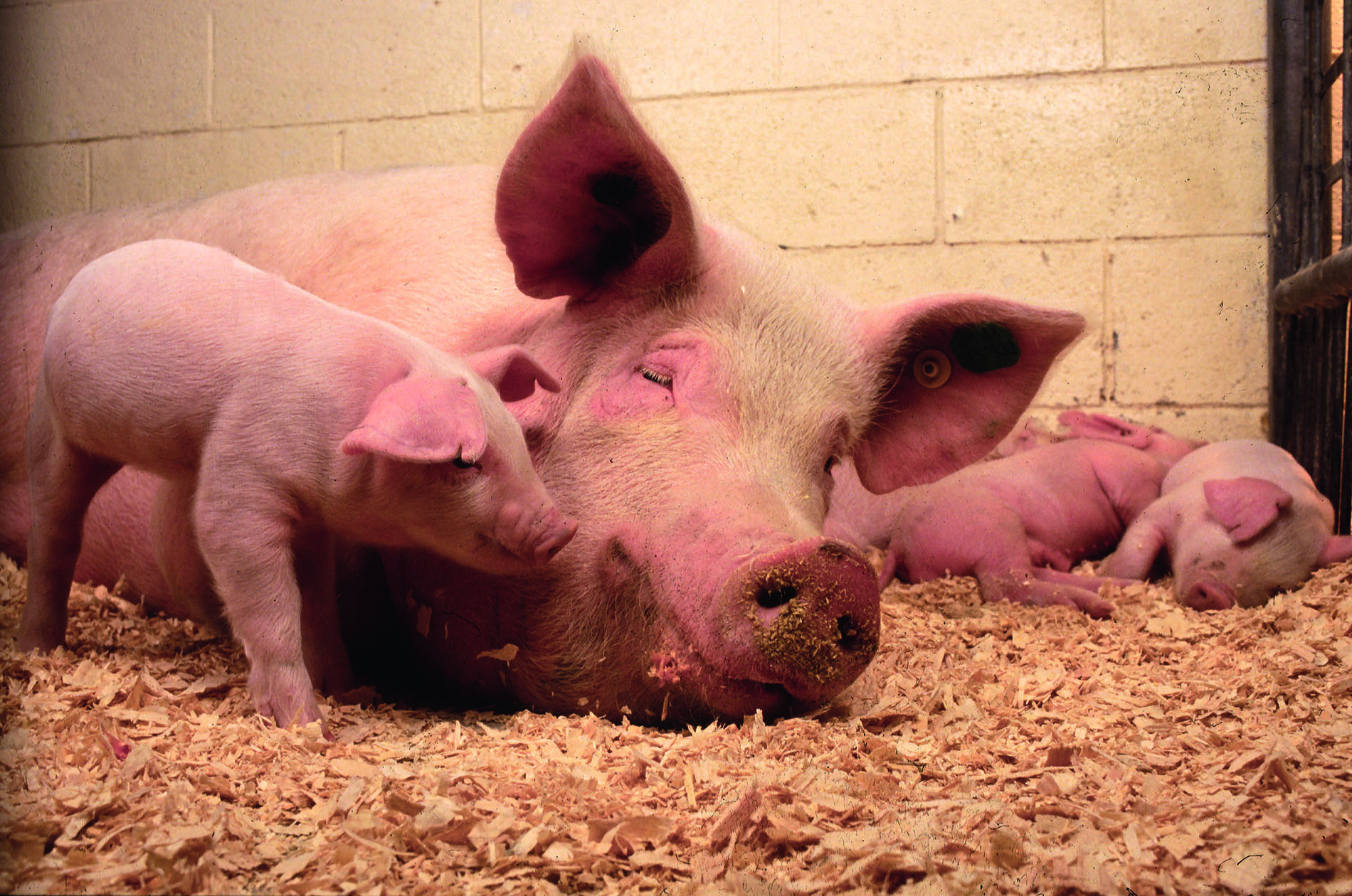
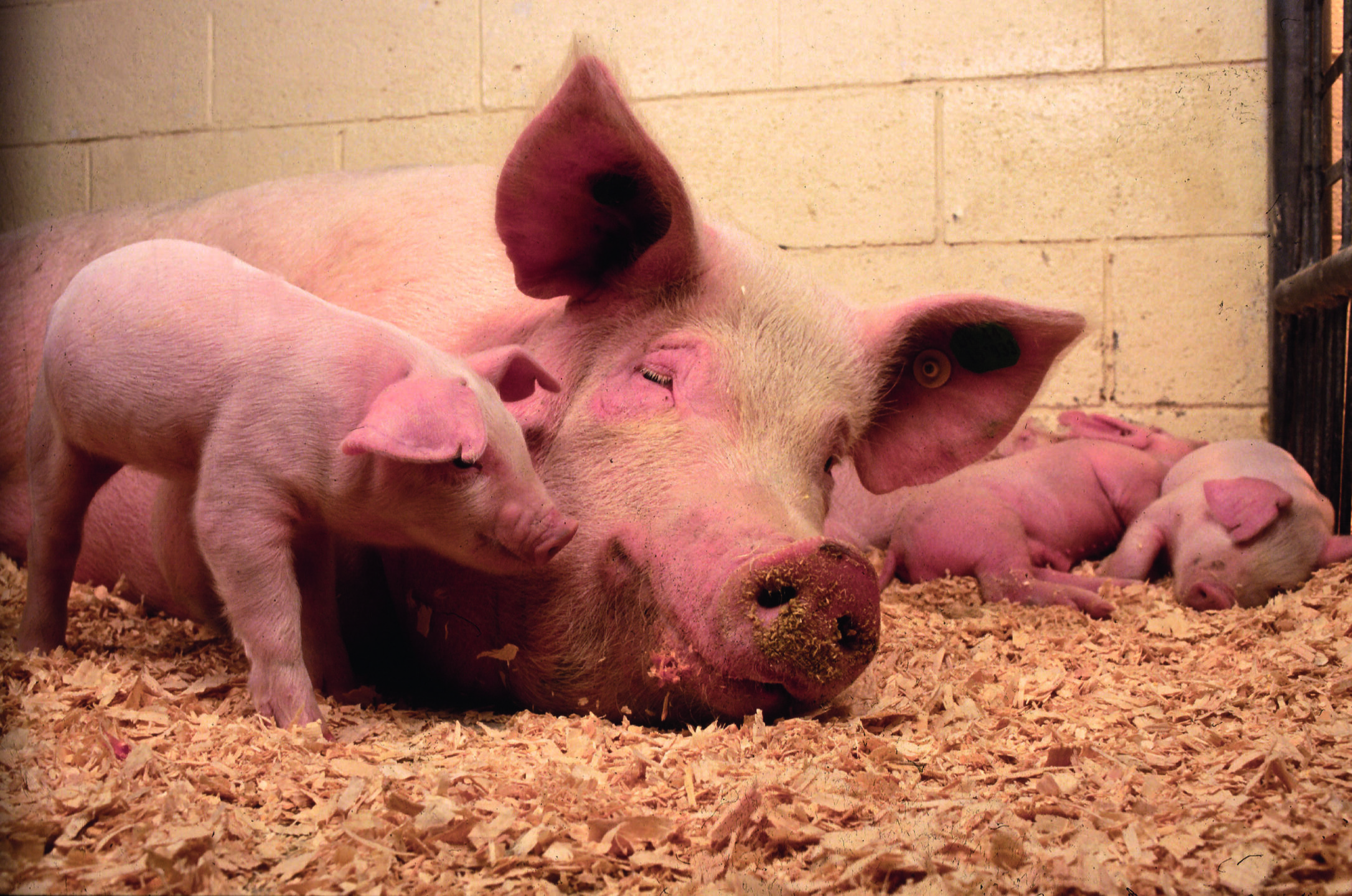
Piglet rearing – there is still room for improvement!

SOW Sau Mit Ferkeln

By I. Heinzl, Editor, and Predrag Persak, Regional Technical Manager North Europe

Optimal rearing conditions for piglets are crucial for ensuring their healthy growth, reducing mortality, and enhancing productivity. These conditions include proper temperature, nutrition, housing, hygiene, and care. Here are the key aspects:

1. Temperature and ventilation

Piglets are sensitive to cold because they cannot regulate their body temperature effectively in the first few days after birth. Proper temperature control is essential to prevent chilling, possibly leading to illness and death. Additionally, regulating the temperature would cost energy, which otherwise could be spent for growth.
Signs of a too-cold environmental temperature are piling on top of one another, tucking the legs under the body, being unable to get up, laying near a corner or wall, or shivering, which may stop if the conditions worsen. Measuring the body temperature shows less than 35°C in the case of chilling.

The following temperatures are recommended for successful piglet rearing:

Farrowing unit (for newborns) 32 – 35°C (90–95°F) during the first few days
After the first week The temperature can gradually decrease by about 1.5-2.0°C per week until it reaches 25°C (77°F)

For supplemental heating, heat lamps, heated floors, or creep areas (a designated warm spot) can be used to maintain the ideal temperature, especially in cooler climates.

Temperature is often closely related to ventilation. Ventilation is essential to reduce dust, humidity, ammonia, and other harmful substances occurring in the air. However, if fresh/cold air enters the pigsty, the temperature decreases, which can get dangerous for the piglets. Suitable ventilation means finding a good balance between providing fresh air and maintaining temperature to prevent energy losses and chilling of the piglets.

Comfort zones can be a solution. They are an effective way to keep the piglets warm and ventilation rates where needed to maintain proper air exchange and humidity levels.

2. Nutrition

Nutrition is critical for piglet growth and immune system development. Most important after birth is the access to colostrum. Piglets are born with an immature immune system, and the maternal antibodies ingested with the colostrum are vital for their survival. They should consume colostrum within the first 6 hours after birth.

It will take 5 to 7 days for piglets to stabilize and get regular on suckling schedule.

At around seven days of age, it is recommended to introduce a highly digestible, nutrient-dense creep feed that helps transition piglets from milk to solid food. Fresh and clean water of the best quality must always be available.

Never forget most important nutrient, beside sow´s love and care – water. Allow piglets free access to the excellent quality water.

3. Housing and Space

A well-designed, clean, and dry environment is critical for reducing stress and promoting health. Farrowing crates help prevent sows from accidentally crushing the piglets during the first few weeks. However, these farrowing crates should provide enough space for the sow to nurse the piglets while allowing piglets to move freely.

Separate warm and clean areas (creep spaces) for the piglets within the farrowing pen are helpful to help the piglets escape from cooler or potentially dangerous parts of the crate. Straw, sawdust, or rubber mats should be provided to keep the piglets warm and comfortable, and good drainage is essential to maintain dryness.

4. Hygiene and Health

Hygiene is crucial to prevent disease and promote the health of piglets. For this purpose, pens and farrowing units should be thoroughly cleaned. Regular removal of waste and keeping bedding dry helps control pathogens. It is essential to clean and disinfect the farrowing unit from one farrowing to the other to reduce disease risks.

Health: After birth, the piglets’ umbilical cord stump should be disinfected to prevent infections. A further essential precautionary measure to prevent anemia is an oral supplementation or an iron injection within the first three days of life, as piglets are born with low iron levels.

For further health monitoring and management, it should be ensured that the piglets are vaccinated against common diseases, such as E. coli, Mycoplasma, and Porcine Circovirus. Additionally, deworming protocols and monitoring for signs of parasites should be implemented for parasite control.

5. Weaning Practices

Piglets are typically weaned between 3 and 4 weeks of age, but early weaning (around 21 days) can be practiced in intensive systems. Optimal weaning requires gradual adaptation to solid feed and a stress-free environment.

If the piglets are weaned at 21 to 28 days, a high-quality starter diet after weaning is essential to maintain growth rates and minimize post-weaning stress.

6. Minimizing Stress

Stress management is essential to prevent disease and poor growth. For this purpose, minimize handling to the minimum during the first few days and, if necessary, handle the piglets gently to reduce stress.

A new environment also means strain for the piglets, so keep the litter groups together during weaning to reduce fighting and social stress.

7. Supportive functional feed ingredients

Depending on veterinary and managing practices, the availability of feed, and the possible use of antimicrobials or other medicals as prophylactics, there can be high variability in rearing conditions in diverse areas of the world. In the following, two functional feed ingredients with entirely different modes of action are presented that support piglets at different rearing conditions.

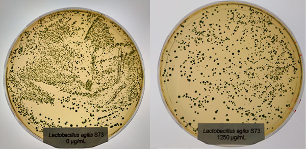
7.1 Egg immunoglobulins (IgY) support piglets under poor rearing conditions

Egg immunoglobulins are beneficial if piglets are not raised under the best conditions, meaning lower hygienic standards and higher pathogenic pressure. With egg immunoglobulins coming from hens having been in contact with pathogens relevant to piglets, it is possible to support the young animals. What is the background? Hens are able to transfer maternal antibodies against diseases that they are confronted with to the egg. With this mechanism, they can provide their progeny with a starter kit for the first time after hatching. However, the best thing is that these antibodies are also helpful for mammals.

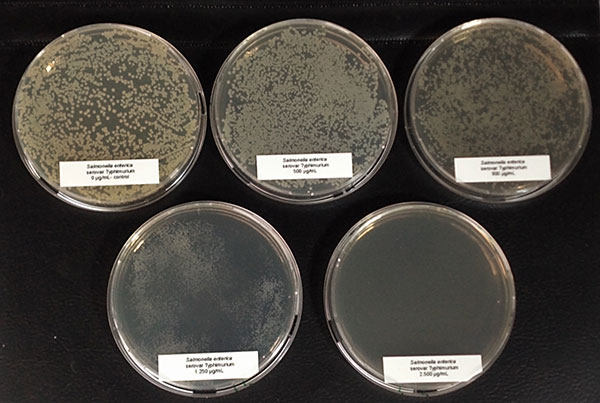
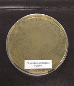
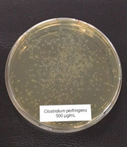
A trial conducted on a commercial farm in Spain shows the weight development of piglets fed an IgY-containing egg powder product (EP) compared to a negative control. The weaned piglets were fed a two-phase feeding (15 days prestarter, 22 days starter). The control (n=51) received no additional functional feed ingredient, whereas the EP group was fed 2 kg of the product/t of feed during the prestarter phase. The animals were weighed individually on days 16 and 37.

The results are shown in Figures 1 and 2.

Figure
Figure 1: Weight development of piglets receiving an IgY-containing egg powder product compared to a negative control
Figure
Figure 2: Daily gain of piglets receiving an IgY-containing egg powder product compared to a negative control

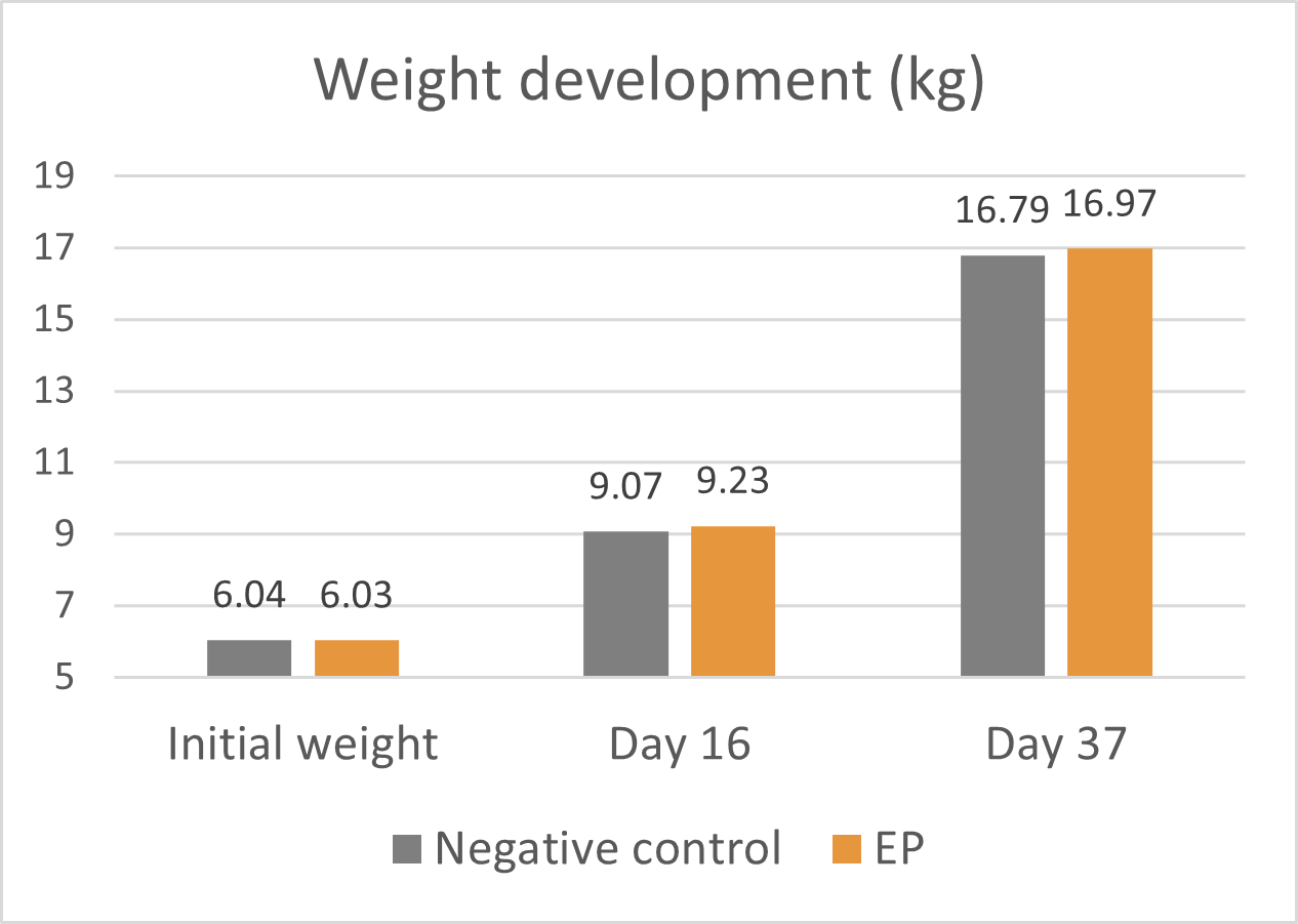
Explanation of the results: Under poor hygienic conditions, the pathogenic pressure is relatively high, and everything lowering this pressure helps to improve gut health, the utilization of nutrients, and performance. Egg immunoglobulins positively influence the gut microbiome, thus helping reduce diarrhea. By lowering the pathogenic pressure, the organism’s energy can be used for growth and must not be employed for the body’s defense.

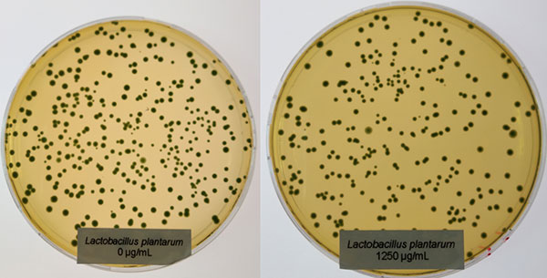
7.2 Phytomolecules can even show improvement under optimum conditions

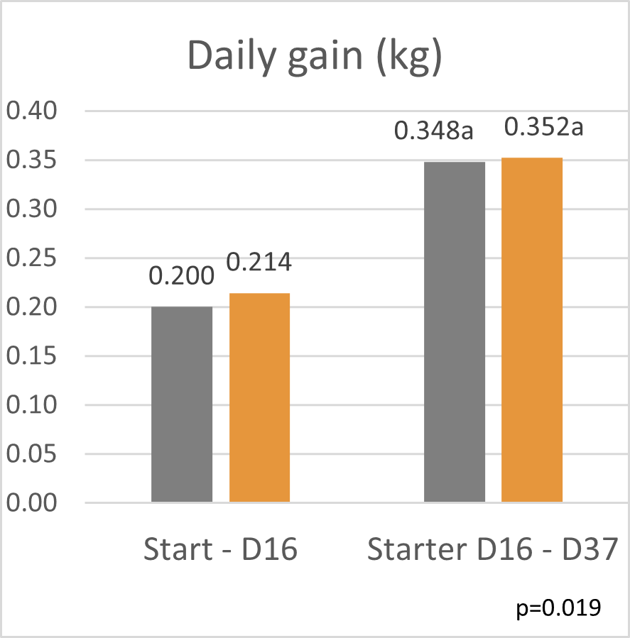
Phytomolecules generally show diverse gut health-promoting effects, from driving the intestinal microbiome in the right direction and strengthening the intestinal barrier to acting as antioxidants or anti-inflammatories or increasing the secretion of digestive juices and, therefore, improving digestion. Which mode of action is relevant if the piglets are raised under already optimal conditions (best hygiene, no prophylactic antibiotics or zinc oxide) and show the highest growth? Is there still room for improvement? Yes, it is. A trial conducted in Germany adduces evidence.

In this trial, 220 piglets weaned on average at 26 days and weighing around 8 kg were housed in 20 pens of 11 castrated males or gilts each. Piglets were blocked by body weight and fed a two-phase feeding program (phase 1 from day 1 to day 13 and phase 2 from day 17 to day 40; pelleted diet). Neither feed or water medication nor therapeutic levels of ZnO were used.

The results of this piglet trial can be seen in Figures 3 and 4.

Figure
Figure 3: Weight development of piglets fed Ventar D compared to a negative control
Figure
Figure 4: Feed conversion rate in piglets fed Ventar D compared to a negative control

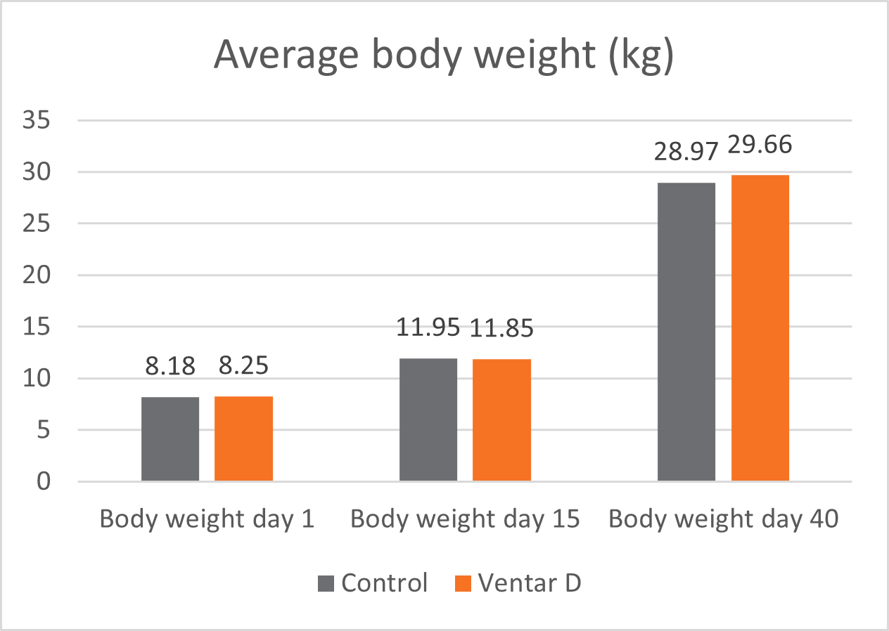
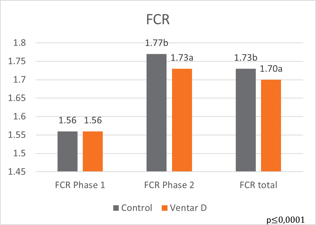
Explanation of the results: The figures show that the piglets in the control already have an extremely high weight compared to those of a similar age in the previous trial, indicating the best rearing conditions in this trial. But, even here, Ventar D has the capacity to improve performance. Why? High-performing animals stress their body more than low-performing ones. Anabolic processes increase oxidative stress and non-infectious inflammation and burden the immune system. The relevant mode of action of Ventar D is not the gut health-promoting or the antimicrobial one because there is no issue. The relevant modes of action in this case are antioxidant and anti-inflammatory. With these two characteristics, Ventar D still has the capacity to improve the performance of piglets that are already at the top level.

8. Conclusion

For high piglet performance, providing the best possible rearing conditions is essential. However, there are differences concerning these conditions in different areas of the world. Depending on them, different feed strategies can be used. Egg immunoglobulins show the best effects if there is a certain pathogenic pressure. Phytomolecules, however, due to their various modes of action, can be beneficial under different levels in rearing conditions. In a low standard, the antimicrobial and gut health-promoting effect is more relevant; in the case of best conditions, the anti-oxidant and anti-inflammatory effects are decisive.

In summary, it could be said that functional feed ingredients have significant advantages in piglet rearing, but the right choice must be made depending on the prevailing conditions.


The crucial role of short-chain fatty acids and how phytomolecules influence them

BROILER PIC Aviagen Drinking Stable

by Dr. Inge Heinzl, Editor EW Nutrition

For optimum health, the content of short-chain fatty acids (SCFAs) is decisive. On the one hand, they act locally in the gut, on the other hand, they are absorbed via the intestinal mucosa into the organism and can affect the whole body. Newer studies in humans show a connection between the deficiency of SCFAs and the occurrence of chronic diseases such as diabetes type 2 or chronic inflammatory gut diseases.

SCFAs – what are they, and where do they come from?

SCFAs consist of a chain of one to six carbon atoms. They are crucial metabolites primarily generated through the bacterial fermentation of dietary fiber (DF) in the hindgut. However, SCFAs and branched SCFAs can also arise during protein fermentation. Short-chain fatty acids predominantly include acetate, propionate, and butyrate, which together account for over 95% of the total SCFAs, typically in a 60:20:20 ratio.

Acetate is produced in two different ways, via the acetyl-CoA and the Wood-Ljungdahl pathways where Bacteroides spp., Bifidobacterium spp., Ruminococcus spp., Blautia hydrogenotrophica, Clostridium spp. are involved. Additionally, acetogenic bacteria can synthesize acetate from carbon dioxide and formate through the Wood-Ljungdahl pathway (Ragsdale and Pierce, 2021). Acetate counts for more than 50% of the total SCFAs in the colon and is the most abundant one.

Propionate can also be produced in two ways. If it is produced via the succinate pathway involving the decarboxylation of methyl malonyl-CoA, the essential bacteria are Firmicutes and Bacteroides. In the acrylate pathway, lactate is converted to propionate. Here, only some bacteria, such as Veillonellaceae or Lachnospiraceae, participate.

Butyrate is produced from acetyl-CoA via the classical pathway by several Firmicutes. However, also other gut microbiota such as Actinobacteria, Proteobacteria, and Thermotogae, which contain essential enzymes (e.g., butyryl coenzyme A dehydrogenase, butyryl-CoA transferase, and butyrate kinase) can be involved. Butyrate can also be produced via the lysine pathway from proteins.

Besides the production of SCFAs from dietary fiber, there is another possibility for the synthesis of SCFAs as well as branched SCFAs – the fermentation of protein in the hindgut. This is something we want to avoid, since it´s clear signal of incorrect animal nutrition. It tells us that there is either oversupply of protein or decrease in protein digestion and absorption.

Which roles do SCFAs play?

SCFAs play a crucial role in the maintenance of gut health. Some benefits originate from these substances’ general character, while others are specific to one acid. If we talk about the benefits of all SCFAs, we can mention the following:

  1. Primarily, SCFAs are absorbed by the intestine and serve enterocytes as an essential substrate for energy production.
  2. By lowering the pH in the intestine, SCFAs inhibit the invasion and colonization of pathogens.
  3. SCFAs can cross bacterial membranes in their undissociated form. Inside the bacterial cell, they dissociate, resulting in a higher anion concentration and bactericidal effect (Van der Wielen et al., 2000)
  4. SCFAs repair the intestinal mucosa
  5. They mitigate intestinal inflammation by G protein-coupled receptors (GPRs).
  6. They enhance immune response by producing cytokines such as IL-2, IL-6, IL-10, and TNF-α in the immune cells. Furthermore, they enhance the differentiation of T-cells into T regulatory cells (Tregs) and bind to receptors (Toll-like receptor, G protein-coupled receptors) on immune cells (Liu et al., 2021).
  7. SCFAs are involved in the modulation of some processes in the gastrointestinal tract, such as electrolyte and water absorption (Vinolo et al., 2011)

After seeing the general characteristics of short-chain fatty acids, let us take a closer look at the specialties of the single SCFAs.

Acetate might play a crucial role in the competitive process between enteropathogens and bifidobacteria and help to build a balanced gut microbial environment (Liu et al., 2021). Additionally, acetate promotes lipogenesis in adipocytes (Liu et al., 2022).

Concerning general health, acetate inhibits, e.g., lung inflammatory response and the reduced air-blood permeability induced by avian pathogenic E. coli-caused chicken colibacillosis (Peng et al., 2021).

Propionate is thought to be involved in controlling intestinal inflammation by regulating the immune cells assisting and, consequently, in maintaining the gut barrier. Furthermore, propionate regulates appetite, controls blood glucose, and inhibits fat deposition in broiler chickens (Li et al., 2021).

In a trial conducted by Elsherif et al. (2022), birds fed a diet with 1.5 g sodium propionate/kg showed considerably (P<0.05) longer and wider guts, higher counts of lactobacillus(P<0.05) and no colonization of Clostridium perfringens. The immunological state improved significantly (P<0.05), which could be seen by the higher antibody titers when the birds were vaccinated against Newcastle disease or avian influenza.

Butyrate additionally improves the function of the intestinal barrier by regulating the assembly of tight junctions (Peng et al., 2009) and stimulating cell renewal and differentiation of the enterocytes. Butyrate-producing microbes on their side prevent the dysbiotic expansion of potentially pathogenic E. coli and Salmonella (Byndloss et al., 2017; Cevallos et al., 2021) by stimulating PPAR-γ signaling. This leads to the suppression of iNOS synthesis and a significant reduction of iNOS and nitrate in the colonic lumen. Furthermore, the microbiota-induced PPAR-γ-signaling inhibits dysbiotic Enterobacteriaceae expansion by limiting the bioavailability of oxygen and, therefore, respiratory electron acceptors to Enterobacteriaceae in the colon.

In a trial conducted by Xiao et al. (2023), sodium butyrate enhanced broiler breeders’ reproductive performance and egg quality due to the regulation of the maternal intestinal barrier and gut microbiota. Additionally, it improved the antioxidant capacity and immune function of the breeder hens and their offspring.

SCFAs’ production can be managed

The extent of production depends on the diet and the composition of the intestinal flora. Nutritional strategies can be taken to regulate the production of short-chain fatty acids by providing dietary fiber and prebiotics, the respective bacteria but also additives in the diet or, on the other, negative way, use of antibiotics.

One example of SCFA-promoting additives is phytomolecules. Ventar D, a blend of diverse gut health-promoting phytomolecules, shows its SCFAs-increasing effect in a trial with Ross 308 broilers.

Trial design: The 41-day research study was conducted at an R&D farm in Turkey, with 3200 Ross 308 broilers in total. The day-old broiler chicks were randomly divided into two groups with 8 replicates in 16-floor pens (6.5×2 m each), each of 200 chicks (100 males and 100 females). One group was managed as a control group with regular feed formulation, and the other group was supplemented with Ventar D. All the birds were provided feeds and water ad libitum. Temperature, lighting, and ventilation were managed as per Ross 308 recommendation.

Groups Application dose
Starter (crumbles) Grower & Finisher – 1 & 2 (pellet)
Control No additive
Ventar D 100 gm/MT 100 gm/MT

All the birds and feed were weighed on days 0, 11, 23, and 41. Dead birds were also weighed, and the feed consumption was corrected accordingly. At the end of the experiment, one male and one female chicken close to the average weight of each pen were separated, weighed, and slaughtered. Short-chain fatty acid (SCFA) concentration in the caecum was measured by gas chromatography (Zhang et al. 2003). Statistical analysis of the data obtained in this study was carried out in the Minitab 18 program using the T-test following the randomized block trial design (P ≤ 0.05). The research results were subjected to statistical analysis on a pen basis. Mortality results were evaluated with the Chi-square test.

Results: Ventar D significantly increased the levels of acetate, butyrate, and total SCFAs. The level of propionate was numerically higher. Additionally, higher final body weights (on average 160 g), improved feed efficiency (6 points), a higher EPEF (33 points), and lower mortality (0.5%) could be asserted in this experiment.

Figure

One explanation could be the microbiota-balancing effect of Ventar D. Meimandipour et al. (2010), for example, saw in their study that increased colonization of Lactobacillus salivarius and Lactobacillus agilis in cecum significantly increased propionate and butyrate formation in caeca.

Phytomolecules: Balancing intestinal microbiome and increasing healthy SCFAs

By promoting beneficial intestinal bacteria and fighting the harmful ones, phytomolecules drive the microbiome in the right direction and promote the production of short-chain fatty acids. Their gut health-protecting effect, in turn, provides for adequate digestion and absorption of nutrients, leading to optimal feed conversion and growth rates. The support of the immune system and the promotion of the antioxidant capacity additionally enhance the health of the animals. Healthy animals grow better, which ultimately leads to a higher profit for the farm.

References:

Byndloss, Mariana X., Erin E. Olsan, Fabian Rivera-Chávez, Connor R. Tiffany, Stephanie A. Cevallos, Kristen L. Lokken, Teresa P. Torres, et al. “Microbiota-Activated PPAR-γ Signaling Inhibits Dysbiotic Enterobacteriaceae Expansion.” Science 357, no. 6351 (August 11, 2017): 570–75. https://doi.org/10.1126/science.aam9949.

Cevallos, Stephanie A., Jee-Yon Lee, Eric M. Velazquez, Nora J. Foegeding, Catherine D. Shelton, Connor R. Tiffany, Beau H. Parry, et al. “5-Aminosalicylic Acid Ameliorates Colitis and Checks Dysbiotic Escherichia Coli Expansion by Activating PPAR-γ Signaling in the Intestinal Epithelium.” mBio 12, no. 1 (February 23, 2021). https://doi.org/10.1128/mbio.03227-20.

Elsherif, Hany M.R., Ahmed Orabi, Hussein M.A. Hassan, and Ahmed Samy. “Sodium Formate, Acetate, and Propionate as Effective Feed Additives in Broiler Diets to Enhance Productive Performance, Blood Biochemical, Immunological Status, and Gut Integrity.” Advances in Animal and Veterinary Sciences 10, no. 6 (June 2022): 1414–22.

Li, Haifang, Liqin Zhao, Shuang Liu, Zhihao Zhang, Xiaojuan Wang, and Hai Lin. “Propionate Inhibits Fat Deposition via Affecting Feed Intake and Modulating Gut Microbiota in Broilers.” Poultry Science 100, no. 1 (January 2021): 235–45. https://doi.org/10.1016/j.psj.2020.10.009.

Liu, Lixuan, Qingqing Li, Yajin Yang, and Aiwei Guo. “Biological Function of Short-Chain Fatty Acids and Its Regulation on Intestinal Health of Poultry.” Frontiers in Veterinary Science 8 (October 18, 2021). https://doi.org/10.3389/fvets.2021.736739.

Liu, Lixuan, Qingqing Li, Yajin Yang, and Aiwei Guo. “Biological Function of Short-Chain Fatty Acids and Its Regulation on Intestinal Health of Poultry.” Frontiers in Veterinary Science 8 (October 18, 2021). https://doi.org/10.3389/fvets.2021.736739.

Meimandipour, A., M. Shuhaimi, A.F. Soleimani, K. Azhar, M. Hair-Bejo, B.M. Kabeir, A. Javanmard, O. Muhammad Anas, and A.M. Yazid. “Selected Microbial Groups and Short-Chain Fatty Acids Profile in a Simulated Chicken Cecum Supplemented with Two Strains of Lactobacillus.” Poultry Science 89, no. 3 (March 2010): 470–76. https://doi.org/10.3382/ps.2009-00495.

Peng, Lu-Yuan, Hai-Tao Shi, Zi-Xuan Gong, Peng-Fei Yi, Bo Tang, Hai-Qing Shen, and Ben-Dong Fu. “Protective Effects of Gut Microbiota and Gut Microbiota-Derived Acetate on Chicken Colibacillosis Induced by Avian Pathogenic Escherichia Coli.” Veterinary Microbiology 261 (October 2021): 109187. https://doi.org/10.1016/j.vetmic.2021.109187.

Peng, Luying, Zhong-Rong Li, Robert S. Green, Ian R. Holzmanr, and Jing Lin. “Butyrate Enhances the Intestinal Barrier by Facilitating Tight Junction Assembly via Activation of AMP-Activated Protein Kinase in Caco-2 Cell Monolayers.” The Journal of Nutrition 139, no. 9 (September 2009): 1619–25. https://doi.org/10.3945/jn.109.104638.

Ragsdale, Stephen W., and Elizabeth Pierce. “Acetogenesis and the Wood–Ljungdahl Pathway of CO2 Fixation.” Biochimica et Biophysica Acta (BBA) – Proteins and Proteomics 1784, no. 12 (December 2008): 1873–98. https://doi.org/10.1016/j.bbapap.2008.08.012.

Vinolo, Marco A.R., Hosana G. Rodrigues, Renato T. Nachbar, and Rui Curi. “Regulation of Inflammation by Short Chain Fatty Acids.” Nutrients 3, no. 10 (October 14, 2011): 858–76. https://doi.org/10.3390/nu3100858.

Wielen, Paul W. van der, Steef Biesterveld, Servé Notermans, Harm Hofstra, Bert A. Urlings, and Frans van Knapen. “Role of Volatile Fatty Acids in Development of the Cecal Microflora in Broiler Chickens during Growth.” Applied and Environmental Microbiology 66, no. 6 (June 2000): 2536–40. https://doi.org/10.1128/aem.66.6.2536-2540.2000.

Xiao, Chuanpi, Li Zhang, Bo Zhang, Linglian Kong, Xue Pan, Tim Goossens, and Zhigang Song. “Dietary Sodium Butyrate Improves Female Broiler Breeder Performance and Offspring Immune Function by Enhancing Maternal Intestinal Barrier and Microbiota.” Poultry Science 102, no. 6 (June 2023): 102658. https://doi.org/10.1016/j.psj.2023.102658.


Oxidative & Inflammatory stress in reproductive Sows

SOW Sau Mit Ferkeln

By Twan van Gerwe, DVM PhD Dipl ECPVS, Technical Director
Dr. Inge Heinzl, Editor


One of the biggest challenges in swine production is keeping the modern, hyperprolific sow healthy and in good shape so that she can wean large, healthy litters and maintain her high reproductive performance.

Unfortunately, sows often suffer from stress and increased systemic inflammation around farrowing and during lactation. This leads to impaired feed intake and disturbed endocrine homeostasis, negatively affecting reproductive and litter performance.

The key to increasing the efficiency of pig production is to reduce the metabolic burden of sows while maintaining the reproductive performance of high-yield sows. A deep understanding of the complex interplay between environmental factors, sow well-being, health, and productivity is necessary to implement enhanced nutritional regimens and meticulous management practices.

Why does oxidative stress occur in today’s sows?

Nowadays, hyperprolific sows produce between 30 and 40 weaned piglets per year and are at a higher risk of suffering from stress. What are the reasons?

A high number of piglets causes oxidative stress

Oxidative stress occurs when reactive oxygen species (ROS) are produced faster than the body’s antioxidant mechanisms can neutralize them and cause damage to lipids, proteins, and DNA. During gestation, the sow needs high amounts of energy to provide for the fetuses. This energy is produced in the placental mitochondria. The placenta, therefore, is a place of active oxygen metabolism during gestation and a source of oxidative stress. In hyperprolific sows, a higher number of fetuses need even more energy to grow. Consequently, ROS production and the risk for intrauterine growth retardation (IUGR) increases (Figure 1). Moreover, evidence shows that the body’s antioxidant potential is reduced in late gestation and after parturition (Szczubial, 2010), resulting in increased oxidative stress biomarkers (Yang, 2023). Increased milk production for large litters demands a substantial amount of energy, risking similar oxidative distress. Therefore, both the final phase of gestation and the subsequent lactation period are predestined for oxidative stress, which has been demonstrated by reduced TEAC (Trolox equivalent antioxidant capacity) levels during these phases (Lee et al., 2023).

SOW Oxidative Stress On Fetus

Figure 1. Illustration of the effect of oxidative stress on the fetus: intrauterine growth retardation (IUGR) (adapted from Yang et al., 2023)

Heat and ambient stress also contribute

The reproductive sow produces lots of heat.  From the beginning of gestation, the sow’s thermoneutral zone decreases. This, however, does not always correspond with the ambient conditions. Especially during the last days of gestation, the discrepancy is exceptionally high as everything is prepared for the newborn piglets, which need a temperature of about 27-35°C. The sow, on the contrary, would be happy with 18-22°C. Additionally, changes around farrowing – moving to the farrowing unit, social stress, change of feed, and the preparation for parturition – exert additional stress for the sows.

Why does the inflammation level increase?

After parturition, systemic inflammation is a normal phenomenon: the reproductive organs have sustained injuries during the parturition process and require remodeling. Inflammation is a natural and desired process, to repair the tissues and return to a normal status. However, inflammation is increased in modern sows, adversely affecting their inflammatory balance. Some possible underlying reasons are:

  1. The high numbers of piglets need a lot of space in the uterus, often leading to damage of the uterine tissue and an inflammatory response in the sows. Lee et al. (2023) found significantly (p<0.10) higher TNF-α concentrations in sows with litters of 15-20 piglets than in sows with 7-14 piglets. TNF-α is a biomarker of inflammation.
  2. Pathogenic infections – particularly infections of the reproductive tract – can induce a prolonged or excessive inflammatory state. A further reason can be the need for more obstetric interventions in hyperprolific sows, which can injure the birth canal or the uterus.
  3. Imbalanced nutrition: Excessive backfat is associated with a higher expression of proinflammatory cytokines, and feed contaminated with mycotoxins can impair the sow’s immunocompetence.

Biomarkers can inform us about the oxidative status

Biomarkers are naturally occurring molecules that help us identify diseases or physiological processes. They provide insights into the oxidative state and inflammatory processes.

Anti-oxidative biomarkers

To check the anti-oxidative capacity, the “beneficial” substances, or antioxidants, can be quantified. These substances can neutralize free radicals or be neutralized by them. Higher levels of antioxidants indicate better antioxidant capacity; when antioxidants are abundant, fewer oxidizable substances have undergone oxidation.

Examples of antioxidant biomarkers:

Total Antioxidant Capacity (T-AOC): represents the synergistic interaction effects of all antioxidants in a matrix (E.g., diet or body fluids). It’s a global measure of non-enzymatic antioxidant efficiency. Various assays, like Trolox Equivalent Antioxidant Capacity (TEAC), which measures a substance’s antioxidant capacity compared to Trolox, can measure T-AOC.

Glutathione Peroxidase (GSH-Px) belongs to the peroxidase family and converts hydrogen peroxide to water.

Catalase (CAT): scavenges ROS. Its activity can predict oxidative stress.

Superoxide Dismutase (SOD): catalyzes the dismutation of superoxide radicals to oxygen and hydrogen peroxide.

Oxidative biomarkers

Oxidative stress biomarkers, the ‘negative’ substances, can also serve as general biomarkers. These include free radicals with oxidant capacity or intermediate/final oxidation products. Ideally, their levels should be minimized.

Examples of oxidative stress biomarkers:

Thiobarbituric acid reactive substances (TBARS): to measure lipid peroxidation products in cells, tissues, and body fluids.

Reactive oxygen species (ROS) or free radicals: unstable, oxygen-containing molecules that react with other molecules in a cell. They might damage DNA, RNA, and proteins and cause cell death. Hydrogen Peroxide (H₂O₂) is a ROS produced during normal cellular metabolism, which causes oxidative damage at excessive levels.

Malondialdehyde (MDA): a final product of oxidative fat degradation and, therefore, a biomarker for lipid peroxidation.

Pro-inflammatory biomarkers

Like oxidative stress, the interplay between pro- and anti-inflammatory signals helps develop the proper immune response for the appropriate duration.

Examples of Pro-inflammatory biomarkers or molecules produced in the case of inflammation:

  • Plasma Adenosine Deaminase (ADA-1 and ADA-2): involved in immune regulation, with ADA-1 inhibiting pro-inflammatory responses and ADA-2 supporting immune cell functions.
  • Interleukins (IL-1α and IL-1β), IL-6: IL-1α and IL-1β are associated with inflammatory diseases, IL-6: is produced during inflammation and acute-phase response.
  • Tumor Necrosis Factor α (TNF-α): endogenous pyrogen that induces fever and promotes inflammation.
  • C-reactive Protein (CRP): liver-produced acute-phase protein responding to inflammation.

Procalcitonin (PCT) is produced by the liver during infections and helps detect bacterial infections.

Examples of anti-inflammatory substances – the “good ones”:

  • Interleukines – IL-4, IL-10: inhibit the function of the macrophages and act, therefore, anti-inflammatory
  • Cortisol: anti-inflammatory and immune-suppressive
  • ACTH: stimulates the production and release of cortisol

Higher stress or infection level lowers performance in sows and piglets

As mentioned, hyperprolific sows suffer from higher oxidative stress, especially during late gestation, parturition, and lactation. Additionally, systemic inflammation occurs to repair the injured tissues to facilitate the healing of the birth canal and remodeling of the uterus to establish the subsequent pregnancy. To this purpose, an inflammatory cascade, triggered by the injuries due to gestation and parturition, involves the release of critical (pro-inflammatory) mediators such as TNF-α and IL-6, leading to the activation of acute phase proteins.

After triggering inflammatory pathways, anti-inflammatory pathways must also be activated to reestablish homeostasis in the reproductive organs (Serhan & Chiang, 2008). Alterations at the onset of anti-inflammatory pathways and exacerbated activation and maintenance of inflammatory pathways can lead to uncontrolled inflammation and the onset of reproductive disease in sows (Kaiser et al., 2018), as well as reduced feed intake and insufficient milk production, resulting in poorly growing piglets and lower weaning weights or piglets suffering from clinical infectious diseases such as diarrhea. If possibly homeostasis cannot be restored, the sow is at risk of contracting diseases like post-partum dysgalactia syndrome (PPDS), lameness, and impaired fertility.

Targeted use of polyphenols can mitigate inflammation and improve the oxidative status of sows

There are several experiments showing the beneficial effects of natural compounds. Especially polyphenols, disposing of phenyl rings and two or more hydroxyl substituents, are perfect radical scavengers and proven antioxidants (Chen, 2023). Phytogenic substances that have anti-inflammatory effects can be found in the families of polyphenols as well as terpenoids, flavonoids, saponins, and tannins (Bunte et al., 2019; Ge et al., 2022; Ginwala et al., 2019; Santos Passos et al., 2022; Ambreen and Mirza, 2020).

Here are some examples showing the beneficial effects of phytochemicals:

  1. Primiparous sows fed with Moringa oleifera leaf meal, rich in polyphenols, saponins, and tannins, illustrate the potential of phytomolecules: serum levels of T-AOC (total anti-oxidative capacity), were increased in late gestation and during lactation, while MDA was reduced. Additionally, piglets that received Moringa oleifera meal showed the highest serum CAT and SOD activities. The meal significantly decreased the farrowing length and number of stillbirths, while there was an increasing trend in the number of live‐born piglets (Sun et al., 2020).
  2. The polyphenol Daidzein, a member of the class of compounds known as isoflavones (200 mg/kg during gestation), increased the total antioxidant capacity (T-AOC) and the activities of glutathione peroxidase and superoxide dismutase. Additionally, it elevated the level of immunoglobulin G and increased the number of piglets born and born alive per litter (Li et al., 2021).
  3. Glycitein, a polyphenol occurring in the isoflavone fraction of soy products, applied during late gestation and lactation increased the total antioxidant capacity and SOD activity during the first 18 days of lactation and the CAT and GSH-Px activity in mid-lactation. Plasma MDA level was reduced from late gestation to the 18th day of lactation. The enhanced oxidative status of the sow resulted in a higher daily gain of the piglets and a higher weaning weight of the litter (Hu et al., 2015).
  4. Meng et al. (2018) tested Resveratrol (300 mg/kg), a stilbenes polyphenol, in sows from day 20 of gestation until farrowing. They saw noticeably higher GSH-Px, SOD, and CAT activities, as well as lower contents of MDA and H2O2 in the placental tissue, improving the antioxidant status of sows and piglets.
  5. Xu et al. (2022) fed silymarin to sows in late gestation. They observed that IL-1ß concentration in the blood sample on the 18th day of lactation was reduced in the supplemented group. The altered fecal microbiota was associated with variations in inflammatory factors, suggesting that silymarin modulates microbiota in the gut and may improve the health of lactation sow.

Phytochemicals support sows against oxidative and inflammatory stress

The above-presented examples show that phytochemicals, particularly those developed to have a potent anti-inflammatory and anti-oxidative capacity, have a high potential to alleviate oxidative stress in pregnant and lactating sows and reduce inflammation when applied in sow diets. Consequently, a broader use of these natural substances should be considered to reduce the metabolic burden of sows and increase the efficiency of pig production.

References:

Ambreen, Madieha, and Safdar Ali Mirza. “Evaluation of Anti-Inflammatory and Wound Healing Potential of Tannins Isolated from Leaf Callus Cultures of Achyranthes Aspera and Ocimum Basilicum.” Pak J Pharm Sci . 33, no. 1 (January 2020): 361–69.

Bunte, Kübra, Andreas Hensel, and Thomas Beikler. “Polyphenols in the Prevention and Treatment of Periodontal Disease: A Systematic Review of in Vivo, Ex Vivo and in Vitro Studies.” Fitoterapia 132 (January 2019): 30–39. https://doi.org/10.1016/j.fitote.2018.11.012.

Chen, Jun, Zhouyin Huang, Xuehai Cao, Tiande Zou, Jinming You, and Wutai Guan. “Plant-Derived Polyphenols in Sow Nutrition: An Update.” Animal Nutrition 12 (March 2023): 96–107. https://doi.org/10.1016/j.aninu.2022.08.015.

Ge, Jiamin, Zhen Liu, Zhichao Zhong, Liwei Wang, Xiaotao Zhuo, Junjie Li, Xiaoying Jiang, Xiang-Yang Ye, Tian Xie, and Renren Bai. “Natural Terpenoids with Anti-Inflammatory Activities: Potential Leads for Anti-Inflammatory Drug Discovery.” Bioorganic Chemistry 124 (July 2022): 105817. https://doi.org/10.1016/j.bioorg.2022.105817.

Ginwala, Rashida, Raina Bhavsar, De Gaulle Chigbu, Pooja Jain, and Zafar K. Khan. “Potential Role of Flavonoids in Treating Chronic Inflammatory Diseases with a Special Focus on the Anti-Inflammatory Activity of Apigenin.” Antioxidants 8, no. 2 (February 5, 2019): 35. https://doi.org/10.3390/antiox8020035.

Hu, Y. J., K. G. Gao, C. T. Zheng, Z. J. Wu, X. F. Yang, L. Wang, X. Y. Ma, A. G. Zhou, and Z. J. Jiang. “Effect of Dietary Supplementation with Glycitein during Late Pregnancy and Lactation on Antioxidative Indices and Performance of Primiparous Sows1.” Journal of Animal Science 93, no. 5 (May 1, 2015): 2246–54. https://doi.org/10.2527/jas.2014-7767.

Kaiser, Marianne, Stine Jacobsen, Pia Haubro Andersen, Poul Bækbo, José Joaquin Cerón, Jan Dahl, Damián Escribano, Peter Kappel Theil, and Magdalena Jacobson. “Hormonal and Metabolic Indicators before and after Farrowing in Sows affected with postpartum Dysgalactia Syndrome.” BMC Veterinary Research 14, no. 1 (November 7, 2018). https://doi.org/10.1186/s12917-018-1649-z.

Lee, Juho, Hyeonwook Shin, Janghee Jo, Geonil Lee, and Jinhyeon Yun. “Large Litter Size Increases Oxidative Stress and Adversely Affects Nest-Building Behavior and Litter Characteristics in Primiparous Sows.” Frontiers in Veterinary Science 10 (August 22, 2023). https://doi.org/10.3389/fvets.2023.1219572.

Li, Yan, Guoru He, Daiwen Chen, Bing Yu, Jie Yu, Ping Zheng, Zhiqing Huang, et al. “Supplementing Daidzein in Diets Improves the Reproductive Performance, Endocrine Hormones and Antioxidant Capacity of Multiparous Sows.” Animal Nutrition 7, no. 4 (December 2021): 1052–60. https://doi.org/10.1016/j.aninu.2021.09.002.

Meng, Qingwei, Tao Guo, Gaoqiang Li, Shishuai Sun, Shiqi He, Baojing Cheng, Baoming Shi, and Anshan Shan. “Dietary Resveratrol Improves Antioxidant Status of Sows and Piglets and Regulates Antioxidant Gene Expression in Placenta by Keap1-Nrf2 Pathway and SIRT1.” Journal of Animal Science and Biotechnology 9, no. 1 (April 20, 2018). https://doi.org/10.1186/s40104-018-0248-y.

Santos Passos, Fabiolla Rocha, Heitor Gomes Araújo-Filho, Brenda Souza Monteiro, Saravanan Shanmugam, Adriano Antunes Araújo, Jackson Roberto Almeida, Parimelazhagan Thangaraj, Lucindo José Júnior, and Jullyana de Quintans. “Anti-Inflammatory and Modulatory Effects of Steroidal Saponins and Sapogenins on Cytokines: A Review of Pre-Clinical Research.” Phytomedicine 96 (February 2022): 153842. https://doi.org/10.1016/j.phymed.2021.153842.

Serhan, C N, and N Chiang. “Endogenous Pro‐resolving and Anti‐inflammatory Lipid Mediators: A New Pharmacologic Genus.” British Journal of Pharmacology 153, no. S1 (March 2008). https://doi.org/10.1038/sj.bjp.0707489.

Sun, Jia‐Jie, Peng Wang, Guo‐Ping Chen, Jun‐Yi Luo, Qian‐Yun Xi, Geng‐Yuan Cai, Jia‐Han Wu, et al. “Effect of Moringa Oleifera Supplementation on Productive Performance, Colostrum Composition and Serum Biochemical Indexes of Sow.” Journal of Animal Physiology and Animal Nutrition 104, no. 1 (October 30, 2019): 291–99. https://doi.org/10.1111/jpn.13224.

Szczubiał, M. “Changes in Oxidative Stress Markers in Plasma of Sows during Periparturient Period.” Polish Journal of Veterinary Sciences, March 3, 2020, 185–90. https://doi.org/10.24425/pjvs.2020.132764.

Xu, Shengyu, Xiaojun Jiang, Xinlin Jia, Xuemei Jiang, Lianqiang Che, Yan Lin, Yong Zhuo, et al. “Silymarin Modulates Microbiota in the Gut to Improve the Health of Sow from Late Gestation to Lactation.” Animals 12, no. 17 (August 26, 2022): 2202. https://doi.org/10.3390/ani12172202.

Yang, Xizi, Ruizhi Hu, Mingkun Shi, Long Wang, Jiahao Yan, Jiatai Gong, Qianjin Zhang, Jianhua He, and Shusong Wu. “Placental Malfunction, Fetal Survival and Development Caused by Sow Metabolic Disorder: The Impact of Maternal Oxidative Stress.” Antioxidants 12, no. 2 (February 2, 2023): 360. https://doi.org/10.3390/antiox12020360.


Mitigating Eimeria resistance in broiler production with phytogenic solutions

Header Image

By Dr. Ajay Bhoyar, Global Technical Manager, EW Nutrition

In modern, intensive poultry production, the imminent threat of resistant Eimeria looms large, posing a significant challenge to the sustainability of broiler operations. Eimeria spp., capable of developing resistance to our traditional interventions, has emerged as a pressing global issue for poultry operators. The resistance of Eimeria to conventional drugs, coupled with concerns over drug residue, has necessitated a shift towards natural, safe, and effective alternatives.

Several phytogenic compounds, including saponins, tannins, essential oils, flavonoids, alkaloids, and lectins, have been the subject of rigorous study for their anticoccidial properties. Among these, saponins and tannins in specific plants have emerged as powerful tools in the fight against these resilient protozoa. In the following, we delve into innovative strategies that leverage the potential of these compounds, particularly saponins and tannins, to prevent losses by mitigating the risk of resistant Eimeria in poultry production.

Understanding resistant Eimeria in broiler production

The World Health Organization Scientific Group (World Health Organization, 1965) developed the definition of resistance in broad terms as ‘the ability of a parasite strain to survive and/or to multiply despite the administration and absorption of a drug given in doses equal to or higher than those usually recommended but within the limits of tolerance of the subject’.

The high reproduction rate of Eimeria spp. allows them to evolve quickly and develop resistance to drugs used for their control. Moreover, the resistant strains of Eimeria can persist in the environment due to their ability to form resistant oocysts, leading to the re-infection of animals and further spread of resistant strains.

Resistant Eimeria strains present many challenges in modern poultry farming, significantly impacting overall productivity and economic sustainability. However, one of the primary challenges is the reduced efficacy of traditional anti-coccidial drugs.

Eimeria resistance occurs in different types

There are different possibilities as to why Eimeria are resistant to specific drugs.

Acquired resistance results from heritable decreases in the sensitivity of specific strains and species of Eimeria to drugs over time. There are two types of acquired resistance: partial and complete. These types depend upon the extent of sensitivity lost. There is a direct relationship between the concentration of the drug and the degree of resistance. A strain controlled by one drug dose may show resistance when a lower concentration of the same drug is administered.

Cross-resistance is the sharing of resistance among different compounds with similar modes of action (Abbas et al., 2011). This, however, may not always occur (Chapman, 1997).

Multiple resistance is resistance to more than one drug, even though they have different modes of action (Chapman, 1993).

Natural substances can bring back the efficacy of anticoccidial measures

It was found that if a drug to which the parasite has developed resistance is withdrawn from use for some time or combined with another effective drug, the sensitivity to that drug may return (Chapman, 1997).

Botanicals and natural identical compounds are well renowned for their antimicrobial and antiparasitic activity, so they can represent a valuable tool against Eimeria (Cobaxin-Cardenas, 2018). The mechanisms of action of these molecules include degradation of the cell wall, cytoplasm damage, ion loss with reduction of proton motive force, and induction of oxidative stress, which leads to inhibition of invasion and impairment of Eimeria spp. development (Abbas et al., 2012; Nazzaro et al., 2013). Natural anticoccidial products may provide a novel approach to controlling coccidiosis while meeting the urgent need for control due to the increasing emergence of drug-resistant parasite strains in commercial poultry production (Allen and Fetterer, 2002).

Saponins and Tannins: Nature’s Defense against Eimeria Challenge

Phytogenic solutions, specifically those based on saponins and tannins, have recently surfaced as promising alternatives to mitigate the Eimeria challenge in poultry production. By harnessing the power of these natural compounds, poultry producers can boost the resilience of their flocks against the Eimeria challenge, promoting both the birds’ welfare and the industry’s sustainability.

Saponins are glycosides found in many plants with distinctive soapy characteristics due to their ability to foam in water. In the context of Eimeria, saponins can disrupt the integrity of the parasites’ cell membranes. When consumed, saponins can interfere with the protective outer layer of Eimeria, weakening the parasite and rendering it vulnerable to the host’s immune responses. This disruption impedes the ability of Eimeria to attach to the intestinal lining and reproduce, effectively curtailing the infection.

Tannins are polyphenolic compounds with astringent properties, occurring in various plant parts, such as leaves, bark, and fruits. Choosing the proper tannin at the right level and time is crucial to realize the benefits of tannin-based feed additives.

In the context of Eimeria, tannins exhibit several mechanisms of action. Firstly, they bind to proteins within the parasites, disrupting their enzymatic activities and metabolic processes. This interference weakens Eimeria, hindering its ability to cause extensive damage to the intestinal lining. Secondly, tannins are anti-inflammatory, reducing the inflammation caused by Eimeria infections. Additionally, tannins act as antioxidants, protecting the intestinal cells from oxidative stress induced by the parasite.

When incorporated into broilers’ diets, saponins and tannins create an unfavorable environment for Eimeria, inhibiting their growth and propagation within the host. Moreover, these compounds fortify the broiler’s natural defenses, enhancing its ability to resist Eimeria infections. By leveraging the innate properties of saponins and tannins, the impact of resistant Eimeria strains can effectively be managed and mitigated, fostering healthier flocks and sustainable poultry production.

What is Pretect D?

Pretect D is a unique proprietary blend of phytomolecules, including saponins and tannins, that supports the control of coccidiosis challenges in poultry production. It can be used alone or in combination with coccidiosis vaccines, ionophores, and chemicals as part of a shuttle or rotation program.

FigureFig.1. Key active ingredients of Pretect D

Modes of action of Pretect D

Pretect D exhibits multiple modes of action to optimize gut health during challenging times. Due to its anti-protozoal, anti-inflammatory, immunomodulatory, and antioxidant properties, it

  1. effectively decreases oocyst excretion and disease spread
  2. promotes restoring the mucosal barrier function and improves intestinal morphology
  3. protects the intestinal epithelium from inflammatory and oxidative damage.

The beneficial effects of Pretect D

The beneficial effects of Pretect D’s inclusion in the coccidiosis control program include improving overall gut health and broiler production performance.

In a challenge study with Cobb 500 broiler chicks under a mixed Eimeria inoculum challenge, it was evident that the group receiving Pretect D (@500g/ton) in the feed throughout the 35-day rearing period had less coccidia-caused lesions (D27) than the broilers challenged and fed control diets.

FigureFig. 2: Pretect D reduced coccidia-caused lesions in broilers

In another field study, a traditional anticoccidial program (Starter and Grower I feeds: Narasin + Nicarbazin, Grower II feed: Salinomycin, Finisher/ withdrawal feeds: No anticoccidial) was compared with a program combining anticoccidials with Pretect D (Starter and Grower I feeds: Narasin + Nicarbazin, Grower II and Finisher feeds: Pretect D). The addition of Pretect D significantly reduced OPG count and lowered the coccidiosis lesion score compared to the control (Fig. 3).

Figure
Figure

Pretect D Reduced Broilers Induc

Fig.3. Pretect D reduced broilers’ coccidiosis lesion score and OPG count

Consequently, broilers receiving Pretect D showed better overall production performance.

Figure
Figure
Figure

Pretect D Reduced Broilers Induc

Fig. 4. Overall improved production performance by Pretect D

Pretect D: Application Strategies

The introduction of an effective phytogenic combination in the coccidiosis control program can help mitigate the drug resistance issue. Such a natural anticoccidial solution can be used as a standalone, preferably in less challenging months, as well as in combination with chemicals (shuttle/ rotation) or a coccidiosis vaccine (bio-shuttle), reducing the need for frequent drug use.

Shuttle programs are commonly employed for managing coccidiosis, and they yield a satisfactory level of success. Within these programs, multiple drugs from distinct classes of anticoccidials are administered throughout a single flock. For instance, one class of drug is utilized in the starter feed, another in the grower stage, reverting to the initial class for the finisher diet and concluding with a withdrawal period.

In rotation programs, anticoccidial drugs are alternated between batches rather than within a single batch.

Conclusions

Coccidiosis is considered one of the most economically significant diseases of poultry and the development of anticoccidial resistance has threatened the profitability of the broiler industry. Therefore, regularly monitoring Eimeria species to develop resistance against different anticoccidial groups is crucial to managing resistance and choosing an anticoccidial. It would be rewarding to use an effective phytogenic solution in the coccidiosis control program as a strategic and tactical measure and to focus on such integrated programs for drug resistance management in the future.

References:

Abbas, R.Z., D.D. Colwell, and J. Gilleard. “Botanicals: An Alternative Approach for the Control of Avian Coccidiosis.” World’s Poultry Science Journal 68, no. 2 (June 1, 2012): 203–15. https://doi.org/10.1017/s0043933912000268.

Abbas, R.Z., Z. Iqbal, D. Blake, M.N. Khan, and M.K. Saleemi. “Anticoccidial Drug Resistance in Fowl Coccidia: The State of Play Revisited.” World’s Poultry Science Journal 67, no. 2 (June 1, 2011): 337–50. https://doi.org/10.1017/s004393391100033x.

Allen, P. C., and R. H. Fetterer. “Recent Advances in Biology and Immunobiology ofEimeriaSpecies and in Diagnosis and Control of Infection with These Coccidian Parasites of Poultry.” Clinical Microbiology Reviews 15, no. 1 (January 2002): 58–65. https://doi.org/10.1128/cmr.15.1.58-65.2002.

Chapman, H. D. “Biochemical, Genetic and Applied Aspects of Drug Resistance inEimeriaParasites of the Fowl.” Avian Pathology 26, no. 2 (June 1997): 221–44. https://doi.org/10.1080/03079459708419208.

Chapman, H.D. “Resistance to Anticoccidial Drugs in Fowl.” Parasitology Today 9, no. 5 (May 1993): 159–62. https://doi.org/10.1016/0169-4758(93)90137-5.

Cobaxin-Cárdenas, Mayra E. “Natural Compounds as an Alternative to Control Farm Diseases: Avian Coccidiosis.” Farm Animals Diseases, Recent Omic Trends and New Strategies of Treatment, March 21, 2018. https://doi.org/10.5772/intechopen.72638.

Nazzaro, Filomena, Florinda Fratianni, Laura De Martino, Raffaele Coppola, and Vincenzo De Feo. “Effect of Essential Oils on Pathogenic Bacteria.” Pharmaceuticals 6, no. 12 (November 25, 2013): 1451–74. https://doi.org/10.3390/ph6121451.

Pop, Loredana Maria, Erzsébet Varga, Mircea Coroian, Maria E. Nedișan, Viorica Mircean, Mirabela Oana Dumitrache, Lénárd Farczádi, et al. “Efficacy of a Commercial Herbal Formula in Chicken Experimental Coccidiosis.” Parasites &amp; Vectors 12, no. 1 (July 12, 2019). https://doi.org/10.1186/s13071-019-3595-4.

World Health Organization Technical Report Series No. 296, (1965) pp:. 29.


Respiratory disease – one of the biggest problems in horses

Houser Home Bg

By Judith Schmidt, Product Manager On-Farm Solutions

The respiratory tract in horses is prone to various problems, ranging from allergic reactions and inflammation to severe infections. Respiratory diseases are a constant topic of suffering and irritation in horse breeding and keeping. According to a study published in 2005, respiratory diseases account for about 40 % of all equine internal diseases recorded worldwide (Thein 2005). Through early diagnosis, appropriate treatment, and preventive measures, horse owners can help maintain the respiratory health of their horses and promote their well-being and performance.

The horse’s lung – a high-performance organ

The respiratory tract of our horses is a high-performance system with a large surface, allowing the exchange between the inside of the body and the environment. The lungs enable the gas exchange, i.e., the transfer of oxygen from the air into the horse’s bloodstream and the discharge of CO2. A functioning gas exchange is crucial for the horse to supply its muscles with sufficient oxygen and perform.

Even when resting, a 600-kg horse breathes about 50 to 80 liters of air per minute into its lungs. With increasing load, this value can rise to 2.000 liters per minute at maximum load. If a horse is healthy, it breathes calmly and slowly and takes eight to sixteen deep breaths per minute.

A special mucous membrane covering the entire respiratory tract protects the lungs from harmful influences. When irritated by pathogens or foreign bodies, this mucous membrane generates higher amounts of mucous and transports it toward the mouth cavity with the help of the finest cilia. In this way, most harmful particles are usually trapped quickly, reliably, and, above all, effectively and, if necessary, coughed up before they can even reach the alveoli and cause damage there.

The most common respiratory diseases in horses

Chronic obstructive bronchitis

Chronic obstructive bronchitis is better known as COB or equine asthma. COB is more common in horses regularly kept in dusty or poorly ventilated environments, such as cramped stables or pastures with high mold levels. Inhalation of dust particles and allergens can cause respiratory tract inflammation, leading to coughing, increased mucus expectoration, and breathing difficulties. The clinical picture of COB can vary greatly. From occasional poor performance in show horses to chronic coughing with purulent nasal discharge or significant weight loss.

Tracheitis

Another common respiratory disease in horses is tracheitis, often caused by bacterial or viral infections. Young and older horses and those with a weakened immune system are particularly susceptible to tracheitis. Besides infections, factors such as dust, smoke, or chemicals can also irritate the mucous membrane of the trachea and trigger inflammation.

Hay fever

Hay fever, also known as allergic respiratory disease or rhinitis, is a common condition affecting horses. Known to humans, it is an allergic reaction to certain pollen, molds, or other environmental allergens that are present in the air. Common signs include sneezing, a runny nose, and itchy eyes. However, some horses may also suffer from coughing or respiratory symptoms. Hay fever in horses can occur seasonally, depending on the pollen emerging, and the symptoms may be more severe during spring, summer, or autumn.

Asthma

Asthma in horses, also known as equine asthma or heaves, is a chronic respiratory disease similar to asthma in humans in many ways. The main cause of this disease is hypersensitivity of the respiratory tract to dust, allergens, or mold spores in the horses’ environment.

How to differentiate between respiratory distress and harmless rattling?

Hourser Side Bg

Horse owners know it – the four-legged friends have an impressive range of breathing sounds. But which are harmless, such as the exited trumpeting through the nostrils during a fright, and which could be respiratory disease symptoms?

Diagnosing respiratory problems in horses can be challenging because symptoms are often non-specific signs and similar to several diseases.

Snorting: When horses snort, it is a sign of relaxation. There is usually no cause for concern—quite the opposite.
Snorting at a gallop: Many horses snort rhythmically at a gallop, which is also considered harmless. Snorting is particularly common in thoroughbreds.
Coughing during, e.g., trotting: Occurs so frequently that it is often perceived as usual. But it is not. Coughing is always an alarm signal and can indicate an allergy, asthma, or a viral or bacterial infection.
Whistling when inhaling: In this case, to be on the safe side, a veterinarian should be consulted.

What are the consequences of respiratory disease?

Respiratory disease in horses can have significant economic consequences. If a horse suffers from chronic obstructive bronchitis or another respiratory illness, this can lead to various problems:

  • Veterinary costs increase: Diagnosing and treating respiratory diseases often require veterinary visits, medication, and possibly further examinations such as x-rays or endoscopy.
  • Performance decreases: A horse with respiratory problems may have severely limited performance. It may have difficulty breathing, negatively affecting its athletic performance, equestrian work, or other activities.
  • Downtime: During the treatment or recovery, horses may have to take a break or be taken out of training, resulting in loss of income, especially if the horse was intended for competition or show.
  • Decrease in value: A horse with chronic respiratory problems may lose its value as a sport or breeding horse. The demand for that horse and, therefore, the selling price might decrease.

Early diagnosis and treatment are crucial for containing the economic impact. However, the best strategy is to minimize the risk of respiratory disease by appropriate preventive measures.

Prevention

Preventing cough in horses is considerably important to reduce the incidence and severity of respiratory disease. Several measures can be taken to achieve this goal:

  1. A clean horse stable is crucial: Dust is a common trigger of respiratory symptoms in horses. Removing dust, dirt, and mold spores regularly from the stable and horse boxes can help improve air quality and reduce respiratory stress.
  2. Allow horses to breathe fresh air with efficient pasture management: When possible, horses should have access to fresh pastures. The natural outdoor environment helps horses breathe cleaner air and inhale fewer harmful particles.
  3. Hay feeding should not increase exposure to allergens: The exposure to allergens can be reduced by choosing high-quality, low-dust hay. Moist soaking of the hay before feeding can also help reduce dust levels.
  4. Ventilation ensures air exchange: Appropriate ventilation in the stable is essential to avoid stagnant air and dust accumulation. The use of fans or natural ventilation systems can improve air circulation.
  5. Feed management: High-quality feed free of molds and allergens can reduce the risk of respiratory problems. It is vital to adjust feed rations to the individual needs of each horse.
  6. Supplements support hygiene measures: Supplements can play a positive role in preventing respiratory problems in horses if used selectively and with expert advice.
    • Immune system support: Supplements such as vitamins, minerals, and antioxidants can strengthen the immune system. A healthy immune system helps the horse to better defend itself against infections and inflammation of the respiratory tract.
    • Certain supplements contain ingredients with anti-inflammatory properties, such as omega-3 fatty acids or herbal extracts. They can help alleviate inflammation in the respiratory tract and thus reduce the risk of respiratory problems.
    • Supporting respiratory health: Some supplements on the market have been specially designed to support respiratory function. They help regulate mucus production, improve respiratory protection, and facilitate the expectoration of mucus.
    • Strengthening lung capacity: Certain ingredients in supplements can support the horse’s lung capacity and promote better oxygen uptake, which is essential for performance and respiratory health.

Conclusion

Respiratory health is essential for horses. So, you should consult the vet in case of noticeable breathing sounds, coughing, fever, or a drop in performance. Respiratory diseases tend to become chronic and long-term problems if they are not treated appropriately. Fresh air and species-appropriate husbandry, feeding dust- and mold-free feed are the first steps to support the normal function of your horse’s respiratory tract. A holistic approach to equine health, including proper stable and feed hygiene, sufficient exercise, and good air quality in the stable is crucial. Appropriate feed supplements can be an excellent tool to round this approach off.

References:

Handbuch Pferd: Dr. med. vet. Peter Thein, 2005

Tierklink Kaufungen (2016): Chronische Obstruktive Bronchitis (COB), Barbara Liese & Dr. Kristian Sander


The future of coccidiosis control

DSC

By Madalina Diaconu, Product Manager Pretect D, EW Nutrition and
Twan van Gerwe, Ph.D., Technical Director, EW Nutrition

With costs of over 14 billion USD per year (Blake, 2020), coccidiosis is one of the most devastating enteric challenges in the poultry industry. With regard to costs, subclinical forms of coccidiosis account for the majority of production losses, as damage to intestinal cells results in lower body weight, higher feed conversion rates, lack of flock uniformity, and failures in skin pigmentation. This challenge can only be tackled, if we understand the basics of coccidiosis control in poultry and what options producers have to manage coccidiosis risks.

Current strategies show weak points

Good farm management, litter management, and coccidiosis control programs such as shuttle and rotation programs form the basis for preventing clinical coccidiosis. More successful strategies include disease monitoring, strategic use of coccidiostats, and increasingly coccidiosis vaccines. However, the intrinsic properties of coccidia make these parasites often frustrating to control. Acquired resistance to available coccidiostats is the most difficult and challenging factor to overcome.

Optimally, coccidiosis control programs are developed based on the farm history and the severity of infection. The coccidiostats traditionally used were chemicals and ionophores, with ionophores being polyether antibiotics. To prevent the development of resistance, the coccidiostats were used in shuttle or rotation programs, at which in the rotation program, the anticoccidial changes from flock to flock, and in the shuttle program within one production cycle (Chapman, 1997).

The control strategies, however, are not 100% effective. The reason for that is a lack of diversity in available drug molecules and the overuse of some molecules within programs. An additional lack of sufficient coccidiosis monitoring and rigorous financial optimization often leads to cost-saving but only marginally effective solutions. At first glance, they seem effective, but in reality, they promote resistance, the development of subclinical coccidiosis, expressed in a worsened feed conversion rate, and possibly also clinical coccidiosis.

Market requests and regulations drive coccidiosis control strategies

Changing coccidiosis control strategies has two main drivers: the global interest in mitigating antimicrobial resistance and the consumer’s demand for antibiotic-free meat production.

Authorities have left ionophores untouched

Already in the late 1990s, due to the fear of growing antimicrobial resistance, the EU withdrew the authorization for Avoparcin, Bacitracin zinc, Spiramycin, Virginiamycin, and Tylosin phosphate, typical growth promoters, to “help decrease resistance to antibiotics used in medical therapy”. However, ionophores, being also antibiotics, were left untouched: The regulation (EC) No 1831/2003 [13]of the European Parliament and the Council of 22 September 2003 clearly distinguished between coccidiostats and antibiotic growth promoters. Unlike the antibiotic growth promoters, whose primary action site is the gut microflora, coccidiostats only have a secondary and residual activity against the gut microflora. Furthermore, the Commission declared in 2022 that the use of coccidiostats would not presently be ruled out “even if of antibiotic origin” (MEMO/02/66, 2022) as “hygienic precautions and adaptive husbandry measures are not sufficient to keep poultry free of coccidiosis” and that “modern poultry husbandry is currently only practicable if coccidiosis can be prevented by inhibiting or killing parasites during their development”. In other words, the Commission acknowledged that ionophores were only still authorized because it believed there were no other means of controlling coccidiosis in profitable poultry production.

Consumer trends drove research on natural solutions

Due to consumers’ demand for antibiotic-reduced or, even better, antibiotic-free meat production, intensified industrial research to fight coccidiosis with natural solutions has shown success. Knowledge, research, and technological developments are now at the stage of offering solutions that can be an effective part of the coccidia control program and open up opportunities to make poultry production even more sustainable by reducing drug dependency.

Producers from other countries have already reacted. Different from the handling of ionophores regime in the EU, where they are allowed as feed additives, in the United States, coccidiostats belonging to the polyether-ionophore class are not permitted in NAE (No Antibiotics Ever) and RWE (Raised Without Antibiotics) programs. Instead of using ionophores, coccidiosis is controlled with a veterinary-led combination of live vaccines, synthetic compounds, phytomolecules, and farm management. This approach can be successful, as demonstrated by the fact that over 50% of broiler meat production in the US is NAE. Another example is Australia, where the two leading retail store chains also exclude chemical coccidiostats from broiler production. In certain European countries, e.g., Norway, the focus is increasingly on banning ionophores.

The transition to natural solutions needs knowledge and finesse

In the beginning, the transition from conventional to NAE production can be difficult. There is the possibility to leave out the ionophores and manage the control program only with chemicals of different modes of action. More effective, however, is a combination of vaccination and chemicals (bio-shuttle program) or the combination of phytomolecules with vaccination and/or chemicals (Gaydos, 2022).

Coccidiosis vaccination essentials

When it is decided that natural solutions shall be used to control coccidiosis, some things about vaccination must be known:

  1. There are different strains of vaccines, natural ones selected from the field and attenuated strains. The formers show medium pathogenicity and enable a controlled infection of the flock. The latter, being early mature lower pathogenicity strains, usually cause only low or no post-vaccinal reactions.
  2. A coccidiosis program that includes vaccination should cover the period from the hatchery till the end of the production cycle. Perfect application of the vaccines and effective recirculation of vaccine strains amongst the broilers are only two examples of preconditions that must be fulfilled for striking success and, therefore, early and homogenous immunity of the flock.
  3. Perfect handling of the vaccines is of vital importance. For that purpose, the personnel conducting the vaccinations in the hatchery or on the farms must be trained. In some situations, consistent high-quality application at the farm has shown to be challenging. As a result, interest in vaccine application at the hatchery is growing.

Phytochemicals are a perfect tool to complement coccidiosis control programs

As the availability of vaccines is limited and the application costs are relatively high, the industry has been researching supportive measures or products and discovered phytochemicals as the best choice. Effective phytochemical substances have antimicrobial and antiparasitic properties and enhance protective immunity in poultry infected by coccidiosis. They can be used in rotation with vaccination, to curtail vaccination reactions of (non-attenuated) wild strain vaccines, or in combination with chemical coccidiostats in a shuttle program.

In a recent review paper (El-Shall et al., 2022), natural herbal products and their extracts have been described to effectively reduce oocyst output by inhibiting Eimeria species’ invasion, replication, and development in chicken gut tissues. Phenolic compounds in herbal extracts cause coccidia cell death and lower oocyst counts. Additionally, herbal additives offer benefits such as reducing intestinal lipid peroxidation, facilitating epithelial repair, and decreasing Eimeria-induced intestinal permeability.

Various phytochemical remedies are shown in this simplified adaptation of a table from El-Shall et al. (2022), indicating the effects exerted on poultry in connection to coccidia infection.

Bioactive compound Effect
Saponins Inhibition of coccidia:
By binding to membrane cholesterol, the saponins disturb the lipids in the parasite cell membrane. The impact on the enzymatic activity and metabolism leads to cell death, which then induces a toxic effect in mature enterocytes in the intestinal mucosa. As a result, sporozoite-infected cells are released before the protozoa reach the merozoite phase.Support for the chicken:
Saponins enhance non-specific immunity and increase productive performance (higher daily gain and improved FCR, lower mortality rate). They decrease fecal oocyst shedding and reduce ammonia production.
Tannins Inhibition of coccidia:
Tannins penetrate the coccidia oocyst wall and inactivate the endogenous enzymes responsible for sporulation.Support for the chicken:
Additionally, they enhance anticoccidial antibodies’ activity by increasing cellular and humoral immunity.
Flavonoids and terpenoids Inhibition of coccidia:
They inhibit the invasion and replication of different species of coccidia.Support for the chicken:
They bind to the mannose receptor on macrophages and stimulate them to produce inflammatory cytokines such as IL-1 through IL-6 and TNF. Higher weight gain and lower fecal oocyst output are an indication of suppression of coccidiosis.
Artemisinin Inhibition of coccidia:
Its impact on calcium homeostasis compromises the oocyst wall formation and leads to a defective cell wall and, in the end, to the death of the oocyst. Enhancing the production of ROS directly inhibits sporulation and also wall formation and, therefore, affects the Eimeria life cycle.Support for the chicken:
Reduction of oocyst shedding
Leaf powder of Artemisia annua Support for the chicken:
Protection from pathological symptoms and mortality associated with Eimeria tenella infection. Reduced lesion score and fecal oocyst output.
The leaf powder was more efficient than the essential oil, which could be due to a lack of Artemisinin in the oil, and to the greater antioxidant ability of A. annua leaves than the oil.
Phenols Inhibition of coccidia:
Phenols change the cytoplasmic membrane’s permeability for cations (H+ and K+), impairing essential processes in the cell. The resulting leakage of cellular constituents leads to water unbalance, collapse of the membrane potential, inhibition of ATP synthesis, and, finally, cell death. Due to their toxic effect on the upper layer of mature enterocytes of the intestinal mucosa, they accelerate the natural renewal process, and, therefore, sporozoite-infected cells are shed before the coccidia reaches the merozoite phase.

Table 1: Bioactive compounds and their anticoccidial effect exerted in poultry

Consumers vote for natural – phytochemicals are the solution

Due to still rising antimicrobial resistance, consumers push for meat production without antimicrobial usage. Phytomolecules, as a natural solution, create opportunities to make poultry production more sustainable by reducing dependency on harmful drugs. With their advent, there is hope that antibiotic resistance can be held in check without affecting the profitability of poultry farming.


Coccidiosis management without increasing antimicrobial resistance – it’s up to us

Header POULTRY BROILERS Broilers

By Tingting Fan, Regional Technical Manager Poultry, EW Nutrition

Chicken coccidiosis is a common and important disease in poultry production, with an incidence of infection as high as 50-70%. The mortality rates are around 20-30% or higher in highly severe cases. In addition to losses due to mortality, producers lose money due to poor growth as well as decreased meat yield and quality. Additionally, the birds get more susceptible to secondary infections, e.g., necrotic enteritis (Moore, 2016).

The costs caused by coccidiosis in poultry are about 13 billion US $ (Blake, 2020). These costs globally divide into 1 billion costs for prophylaxis/treatment and 12 billion due to performance losses. Until now, only 5% of the prophylaxis costs have been created by natural solutions. That means that there is still a high potential to be tapped.

Natural solutions, unfortunately, are only used by a minority

For a long time, ionophores fitting the classical definition of antibiotics and chemicals were used in coccidia-fighting programs – and contributed to the development of antimicrobial resistance (Nesse et al., 2015). Nowadays, the combination with vaccination in rotation or shuttle programs has reduced this danger, but there is still potential. Meanwhile, some natural solutions are available that can be integrated into coccidiosis-fighting programs. However, producers using natural solutions are still a minority.

For thousands of years, plants have been used in human and veterinary medicine. Before the discovery of antibiotics in 1928, diseases were fought with plants. To regain the effectiveness of antibiotics, using natural solutions for prophylaxis should be once more standard, and the use of antibiotics is the treatment only for critical cases.

How does Eimeria damage broilers

The pathogenic mechanism of coccidia or Eimeria spp. is mainly the massive destruction of host intestinal cells when it reproduces, resulting in severe damage to the intestinal mucosa. On the one hand, the damaged gut wall loses its capability for effective digestion and absorption of nutrients, leading to worse feed conversion and lower weight gain.

On the other hand, this damage reduces the chicken’s immunity and paves the way for other infections, such as necrotic enteritis, and raises mortality.

Table 1:The seven most known Eimeria species in broilers and their main site of occurrence

Eimeria species Predilection site
E. tenella Ceca
E. acervulina Duodenum and prox. jejunum
E. maxima Central jejunum
E. mitis Distal jejunum and ileum
E. necatrix Central jejunum and ceca
E. brunetti Ileum, entrance of the ceca and rectum
E. praecox Duodenum and prox. jejunum

Concerning their pathogenicity, for poultry, the Eimeria species must be ordered in the following way: E. necatrix> E. tenella > E. brunetti > E. maxima > E. acervulina > Eimeria mitis, and Eimeria praecox.

Prevention is better than treatment

Thanks to its bi-layered wall with a robust structure, the oocysts of coccidia are extremely resilient. They can survive 4 to 9 months in the litter or soil and are resistant to common disinfectants. Farm personnel and visitors are also important vectors, so good biosecurity practices can reduce the number of oocysts contaminating the premises and help prevent clinical out-brakes. Coccidiosis control in poultry should focus on “prevention” rather than “treatment”, combining biosecurity practices, feed additives, and/or vaccination.

Effective hygiene on the farm is crucial

To prevent coccidia infections, one of the most critical points is hygiene. Biosecurity practices are crucial and include cleaning and disinfection of the poultry houses and their surroundings, pest control and prevention, restriction, control, and management of the entry of personnel, visitors, vehicles, and equipment, among others.

Coccidia oocysts are ubiquitous and survive for a long time, and even effective cleaning and disinfection cannot completely remove them. After a severe outbreak, it is recommended to take drastic biosecurity measures such as flame or caustic soda disinfection to prevent further spread of the disease.

When there are birds in the house, it must be paid attention that the litter is not excessively humid. Litter moisture should be maintained around 25%; turning and replacing moist litter are the best practices to follow. For keeping the litter dry, adequate ventilation and appropriate stocking density are beneficial.

To avoid unnecessary stress and gut health issues, the birds must be fed according to their requirements with high-quality feed so that the animals build up good immunity and resilience.

Coccidiosis can be controlled with effective programs

Anticoccidial drugs were the first means of preventing and controlling coccidiosis in chickens and once achieved very good results. Since Sulfaquinoxaline was found to be effective in the 1850s, about fifty other drugs have been developed for the prevention and control of coccidiosis. Generally, the anticoccidials used for years to prevent the disease can be divided into ionophores and chemicals.

Ionophores, produced as by-products of bacterial fermentation, are technically antibiotics. The great benefits of ionophores are that they kill the parasite before it can infect the bird and thus prevent damage to the host cells. Eimeria species also take a long time to develop resistance to ionophores (Chapman, 2015). Well-established ionophores are products that contain monensin, lasalocid, salinomycin, narasin, or maduramycin; the trade names are Coban/Monensin, Avatec, Coxisstac, Monteban, and Cygro.

Chemicals, these molecules, are produced by chemical synthesis. They differ from each other and ionophores as each one has a unique mode of action against coccidia. In general, they act by interfering with one or more stages of the life cycle of Eimeria, e.g., supplying fake nutrients (Amprolium, Vit. B1) to the parasite, starving them out. The active components here are nicarbazin, amprolium, zoalene, decoquinate, clopidol, robenidine and diclazuril, and the respective trade names Nicarb, Amprol, Zoamix, Deccox, Coyden, Robenz and Clinacox. Eimeria species develop resistance to these chemical molecules; therefore, they must be used carefully and with strict planning. However, cross-resistance does not develop, making them highly valuable in rotation programs.

Vaccination against coccidiosis is accepted by many farmers as a good solution to control coccidiosis in chickens. Vaccination aims to replace resistant field strains with vaccine strains, which are sensitive to anticoccidials. Currently, commercial chicken vaccines are available in natural and attenuated strains; research to obtain safer and more efficient vaccines is also ongoing.

Non-attenuated vaccines are less expensive and make for good immunity, but as they may mildly damage the intestinal epithelium, the risk of necrotic enteritis can increase. On the contrary, attenuated strains – usually “precocious” strains with shorter reproduction cycles, cause less intestinal damage and thus have a lower risk of provoking bacterial or necrotic enteritis. The immunity is like after normal infections; however, you have a controlled epidemiology, fewer coccidiosis outbreaks, and an improved uniformity of the flock.

Phytomolecules-based natural anticoccidials saponins and tannins are natural components that can also help control coccidiosis (e.g., Pretect D, EW Nutrition GmbH). These ingredients act in different ways: the tannins improve the intestinal barrier function locally and systemically. The saponins directly impact the oocysts by preventing their growth, interacting with the cholesterol in the cell membrane (triterpenoid saponin), or hindering further sporulation and causing cell death by causing pores in the cell membrane of the parasite. Altogether, Pretect D promotes the beneficial microbial population and reduces the harmful one, improves the gut barrier function, reduces mucosal inflammation, inhibits growth and replication of Eimeria, preventing their lesions, and fosters birds’ immune response against Eimeria spp.

To prove Pretect D’s effectiveness in the reduction of coccidiosis, several trials were conducted. One of the trials was carried out in Poland with 360.000 broilers in commercial conditions. The animals were divided into ten houses, and two cycles were tested. Half of the birds served as control and received Narasin and Nicarbazin in the starter and grower I diet and salinomycin in the grower II diet. The other half also were fed Narasin and Nicarbazin in the starter and grower I diet, but Pretect D @1kg/t in grower II and 0.5kg/t in the finisher diet. The results are shown in figure 1: The application of Pretect D in the grower II and finisher diet decreased the number of oocysts in the droppings more than the application of salinomycin and, therefore, reduced the spreading of coccidiosis. In addition, the performance of the broilers receiving Pretect D was nothing short of the control’s performance showing Pretect as an optimal completion in shuttle or rotation programs (see more HERE).

Figure Reduction Of Oocysts In The Droppings By Pretect DFigure 1: Reduction of oocysts in the droppings by Pretect D

Managing coccidiosis without promoting antimicrobial resistance is not easy, but feasible

Coccidiosis is a challenge aggravated by our current high level of production. Tools such as ionophores, chemicals, but also vaccines, and natural products are available to fight coccidiosis. However, due to the high probability of resistance development, these tools must be used carefully and in structured programs. The phytomolecules-based product Pretect D gives the possibility to reduce antimicrobial resistance as part of programs against coccidiosis.

References upon request


Pathogenic Enterococcus cecorum – an emerging profit killer for broiler producers

Header

By Dr. Ajay Bhoyar, Global Technical Manager, EW Nutrition

Pathogenic Enterococcus cecorum (EC) is emerging as a significant challenge in poultry production worldwide, causing substantial losses to commercial flocks. EC has become a considerable concern for the poultry industry, not only because of its rapid spread and negative impact on broiler health but also because of its increasing antibiotic resistance. As a result, there is a growing need to explore alternative ways of controlling this bacterium. There is no silver bullet yet as a replacement for antibiotics to limit the load of E. cecorum. Maintaining optimum gut health to avoid E. cecorum leakage during the first week of the broiler’s life can control losses due to E. cecorum.

Phytogenic compounds, which are derived from plants, have gained attention in the last decades as a potential solution for controlling common gut pathogens. These natural compounds have been found to possess antimicrobial properties and can help improve gut health in broilers. In this article, we will discuss the current state of E. cecorum and explore potential strategies, including using phytogenic compounds as support in controlling economic losses due to this emerging pathogen in broiler production.

Enterococcus cecorum and its negative impact on broiler production

E. cecorum is a component of normal enterococcal microbiota in the gastrointestinal tract of poultry. These are facultatively anaerobic, gram-positive cocci. Over the past 15 years, pathogenic strains of E. cecorum have emerged as an important cause of skeletal disease in broiler and broiler breeder chickens (Broast et al., 2017; Jackson et al., 2004 and Jung et al., 2017). Along with the commensal strains of EC, the pathogenic strains also occur and can result in Enterococcal spondylitis (ES), also known as “kinky back”, a serious disease of commercial poultry production in which the bacteria translocate from the intestine to the free thoracic vertebrae and adjacent notarium or synsacrum, causing lameness, hind-limb paresis and, in 5 to 15% of cases, mortality (de Herdt et al., 2008; Martin et al., 2011; Jung and Rautenschlein, 2014). The compression of the spinal cord due to infection of the free thoracic vertebra results in the so-called “kinky back” in the skeletal phase of E. cecorum infection. Kinky back is also a common name for spondylolisthesis, a developmental spinal anomaly. EC is normally found in the gastrointestinal tract and may need the help of other factors, such as a leaky gut, to escape the gastrointestinal tract. The emergent pathogenic strains of E. cecorum have developed an array of virulence factors that allow these strains to 1) colonize the gut of birds in the early life period; 2) escape the gut niche; 3) spread systemically while evading the immune system; and 4) colonize the damaged cartilage of the free thoracic vertebra (Borst, 2023). The E. cecorum can invade internal organs and produce lesions in the pericardium, lung, liver, and spleen.

The negative impact of E. cecorum on broiler economics, health, and welfare

Enterococcus cecorum can harm broiler health, welfare, and economics. This can result in decreased profitability for broiler producers.

The broiler flocks infected with E. cecorum may have reduced feed intake/ nutrient absorption and reduced growth rates, leading to a higher feed conversion ratio, longer production cycles, and lower weight gain. The morbidity and mortality from E. cecorum infection can be as high as 35 % and 15%, respectively. The higher condemnations of up to 9.75% at the processing plant can further add to the losses (Jung et al., 2018).  This can result in significant economic losses for producers.

Further, E. cecorum infections can impair the immune function of broilers, making them more susceptible to other pathogens and reducing their overall health and welfare. Pathogenic E. cecorum is an opportunistic pathogen that can gain momentum during coinfection with E. coli and other gut pathogens, causing a leaky gut. Therefore, a holistic approach to gut health management may help reduce the losses.

E Cecorum

Antibiotic resistance in E. cecorum

E. cecorum has been found to be resistant to multiple antibiotics. Multidrug resistant pathogenic E. cecorum could be recovered from lesions in whole birds for sale at local grocery stores (Suyemoto et al., 2017). Antibiotic resistance can make it difficult to treat and control infections in broilers. This can lead to increased use of multiple antibiotics, which can contribute to the development of antibiotic-resistant bacteria and pose a risk to human health.

Transmission of E. cecorum in broiler flocks

Despite the rapid global emergence of this pathogen, and several works on the subject, the mechanism by which pathogenic E. cecorum spreads within and among vertically integrated broiler production systems remains unclear (Jung et. al.2018). The role of vertical transmission of pathogenic E. cecorum remains elusive. Experimentally infected broiler breeders apparently do not pass the bacterium into their eggs or embryos (Thoefner and Peter, 2016). However, it has been noted that a very low frequency of infected chicks can cause a flock-wide outbreak.

Horizontal transmission: E. cecorum can be transmitted between birds within a flock through direct contact or exposure to contaminated feces, feed, or water.

While the mode of transmission between flocks has not been definitively identified, pathogenic E. cecorum demonstrates rapid horizontal transmission within flocks. It can spread rapidly within flocks via fecal-oral transmission.

Personnel and equipment: E. cecorum can be introduced into a flock through personnel or equipment that has been in contact with infected birds or contaminated materials. For example, personnel working with infected flocks or equipment used in infected flocks can spread the pathogen to uninfected flocks.

Symptoms and diagnosis of E. cecorum in broilers

Enterococcus cecorum infections in broilers can present a range of symptoms, from mild to severe. The most common symptom noticed with E. cecorum is paralysis, which is due to an inflammatory mass that develops in the spinal column at the level of the free thoracic vertebra (FTV). Recognition of this spinal lesion has given rise to several disease names for pathogenic E. cecorum infection, which include vertebral osteomyelitis, vertebral enterococcal osteomyelitis and arthritis, enterococcal spondylitis (ES), spondylolisthesis and, colloquially, “kinky-back” (Jung et al. 2018).

E. cecorum infections can exhibit increased mortality due to septicemia in the early growing period. In this sepsis phase, the clinical signs of E. cecorum may include fibrinous pericarditis, perihepatitis, and air-sacculitis. These lesions might be confused with other systemic bacterial infections like colibacillosis. Therefore, a pure culture is needed for the correct diagnosis of E. cecorum.

The second phase of mortality due to dehydration and starvation of the paralyzed birds can be observed during the finisher phase peaking during 5-6 weeks of age. Paralysis from infection of the free thoracic vertebra is the most striking feature of this disease, with affected birds exhibiting a classic sitting position with both legs extended cranially (Brost et al., 2017).

Diagnosis of E. cecorum in broilers can be challenging, as the symptoms of infection can be similar to those of other bacterial or viral infections. However, a combination of clinical signs, post-mortem examination, and laboratory testing can help to confirm the presence of E. cecorum. Laboratory tests such as bacterial culture and polymerase chain reaction (PCR) can be used to identify the pathogen and also to determine its antibiotic susceptibility. Veterinarians and poultry health professionals can work with producers to develop a diagnostic plan and implement appropriate control measures to manage E. cecorum infections in broiler flocks.

E Cecorum

Prevention and Control of Enterococcus cecorum

The broiler producers/ managers should work with their veterinarians and poultry health professionals to develop an integrated approach to control the spread of E. cecorum and prevent its negative impact on broiler health and productivity.

Currently, there is no commercial vaccine available for preventing pathogenic E. cecorum infection. Therefore, controlling Enterococcus cecorum infection in broiler flocks requires a multifaceted approach that addresses the various modes of transmission and bacterial resistance to antibiotics.

Implementing strict biosecurity protocols, such as controlling access to the farm, disinfecting equipment and facilities, and implementing proper hygiene protocols throughout the integrated broiler operation, can help to minimize the risk of transmission.

Thorough washing of trays and chick boxes in the hatchery with hot water (60-62°C) mixed with an effective disinfectant can reduce the possible vertical transmission of E. cecorum. The vertical transmission may also be prevented by adopting the practice of separating the dirty floor eggs from clean hatching eggs and setting them in the lower racks of the incubator.

Generally, pathogenic isolates from poultry were found to be significantly more drug-resistant than commensal strains (Borst et al., 2012). The selection of an effective antibiotic for the treatment of E. cecorum should be made based on the results of the antibiotic sensitivity test. Antibiotic therapy may not help with paralyzed birds, which ultimately need to be culled. Reducing the use of antibiotics and implementing prudent use practices can help to reduce the development of antibiotic resistance in E. cecorum and other bacteria.

Probiotics can help to maintain the balance of the gut microbiota and may have a protective effect against E. cecorum infections. Fernandez et al. (2019) reported the inhibitory activities of proprietary poultry Bacillus strains against pathogenic isolates of E. cecorum in vitro, but effects are highly strain-dependent and vary significantly among different pathogenic isolates.

Phytogenic compounds and organic acids have been shown to have antimicrobial properties. Phytomolecule-based preparations may help to control E. cecorum infections in broiler flocks in the first week of life, reducing the chances of its translocation from the intestine.

Phytomolecules-based liquid formulations for on-farm drinking water application can also be a handy tool to manage gut health challenges, especially during risk periods in the life of broilers. Such liquid phytomolecule preparations can help to quickly achieve the desired concentration of the active ingredients for a faster antimicrobial effect.

However, these alternatives to antibiotics may be effective only when the E. cecorum is still localized within the gut during the first two weeks of the broiler chicken’s life.

Phytomolecules, also known as phytochemicals, are naturally occurring plant compounds that have been found to have antimicrobial properties. Especially for commercial poultry, nutraceuticals such as phytochemicals showed promising effects, improving the intestinal microbial balance, metabolism, and integrity of the gut due to their antioxidant, anti-inflammatory, immune modulating, and bactericidal properties (Estevez, 2015). Phytogenic compounds have been studied for their potential use in controlling gut pathogens in poultry. Here are some of the roles that phytomolecules can play in controlling gut pathogens:

Antimicrobial activity: Several phytomolecules, such as essential oils, flavonoids, and tannins, have been found to have antimicrobial activity.  Hovorková et al (2018) studied the inhibitory effects of hydrolyzed plant oils (palm, red palm, palm kernel, coconut, babassu, murumuru, tucuma, and Cuphea oil) containing medium-chain fatty acids (MCFAs) against Gram-positive pathogenic and beneficial bacteria. They concluded that all the hydrolyzed oils were active against all tested bacteria (Clostridium perfringens, Enterococcus cecorum, Listeria monocytogenes, and Staphylococcus aureus), at 0.14–4.5 mg/ml, the same oils did not show any effect on commensal bacteria (Bifidobacterium spp. and Lactobacillus spp.). However, further research is needed to test the in-vivo efficacy of phytogenic compounds against pathogenic E. cecorum infections in poultry.

Anti-inflammatory activity: The other coinfecting gut pathogens of E. cecorum can cause inflammation in the intestinal tract of poultry. This can lead to reduced feed intake and growth. Some phytomolecules have been found to have anti-inflammatory activity and can reduce the severity of inflammation. Capsaicin, a naturally occurring bioactive compound in chili peppers, was found to have antioxidant and anti-inflammatory activity. The tendency of capsaicin to substantially diminish the release of COX-2 mRNA is thought to be the reason for its anti-inflammatory effects (Liu et al., 2021).  Thyme oil reduced the synthesis and gene expression of TNF-α, IL-1B, and IL-6 in activated macrophages in a dose-dependent manner, with upregulation of IL-10 secretion (Osana and Reglero, 2012). Cinnamaldehyde has been shown to decrease the expression of several cytokines, such as IL-1 β, IL-6, and TNF-α, as well as iNOS and COX-2, in in-vitro studies (Pannee et al., 2014).

Antioxidant activity: Oxidative stress may contribute to the development of E. cecorum infections in poultry. Phytomolecules, such as polyphenols and carotenoids, have been found to have antioxidant activity and can reduce oxidative stress in the gut of poultry, which can help to prevent E. cecorum infections. Polyphenols widely exist in a variety of plants and have been used for various purposes because of their strong antioxidant ability (Crozier et al., 2009). Quercetin, a flavonoid compound widely present in vegetables and fruits, is well-known for its potent antioxidant effects (Saeed et al., 2017).

Phytomolecules can also modulate the immune system of poultry, which can help to prevent E. cecorum infections. For example, some flavonoids and polysaccharides have been found to enhance the immune response of poultry. Fahnani et. al. (2019) found that supplementing broiler chickens with a combination of flavonoids and polysaccharides extracted from the mushroom Agaricus blazei enhanced their immune response.

Overall, phytomolecules have shown promise in supporting the optimum gut health of poultry. Many phytogenic preparations available in the market can be regarded as an important tool to reduce the use of antibiotics in animal production and mitigate the risk of antimicrobial resistance. However, more research is needed to develop an effective combination of active ingredients, as well as strategies for their use in controlling E. cecorum infections in poultry.

Conclusion

In conclusion, the emergence of pathogenic strains of E. cecorum is becoming a major concern for broiler producers globally. This bacterial pathogen can cause significant economic losses in the broiler industry by affecting the overall health and productivity of the birds. Pathogenic E. cecorum infection can lead to clinical signs including diarrhea, decreased feed intake, reduced growth rate, and increased mortality. Proactive measures must be taken to prevent the introduction and spread of pathogenic E. cecorum in broiler flocks. Implementing strict biosecurity protocols and proper disinfection procedures can help reduce the risk of E. cecorum infection. The use of effective antibiotics after receiving the results of the antibiotics sensitivity test is a crucial step in controlling the infection. Phytomolecule-based preparations can be a potential alternative to control the load of E. cecorum by maintaining optimum gut health to minimize economic losses. Moreover, ongoing surveillance and monitoring of pathogenic E. cecorum prevalence in the broiler industry can assist in the timely detection and control of outbreaks.

In summary, the emergence of pathogenic E. Cecorum as a profit killer in the broiler industry warrants careful attention and proactive management practices to minimize its impact.

References:

Borst, L. B., M. M. Suyemoto, A. H. Sarsour, M. C. Harris, M. P. Martin, J. D. Strickland, E. O. Oviedo, and H. J. Barnes. “Pathogenesis of Enterococcal Spondylitis Caused by Enterococcus cecorum in Broiler Chickens”. Vet. Pathol. 54:61-73. 2017.

Crozier A., Jaganath I.B., Clifford M.N. “Dietary phenolics: Chemistry, bioavailability and effects on health”. Nat. Prod. Rep. 2009;26:1001–1043.

De Herdt, P., P. Defoort, J. Van Steelant, H. Swam, L. Tanghe, S. Van Goethem, and M.

593 Vanrobaeys. “Enterococcus cecorum osteomyelitis and arthritis in broiler chickens”. Vlaams Diergeneeskundig Tijdschrift 78:44-48. 2009.

Estévez, M. “Oxidative damage to poultry: From farm to fork”. Poult. Sci. 2015, 94, 1368–1378.

Fanhani, Jamile & Murakami, Alice & Guerra, Ana & Nascimento, Guilherme & Pedroso, Raíssa & Alves, Marília. (2016). “Agaricus blazei in the diet of broiler chickens on immunity, serum parameters and antioxidant activity”. Semina: Ciencias Agrarias. 37. 2235-2246.

Hovorková P., Laloučková K., Skřivanová E. (2018): “Determination of in vitro antibacterial activity of plant oils containing medium-chain fatty acids against Gram-positive pathogenic and gut commensal bacteria”. Czech J. Anim. Sci., 63: 119-125.

Jung, A., and S. Rautenschlein. “Comprehensive report of an Enterococcus cecorum infection in a broiler flock in Northern Germany. BMC Vet. Res. 10:311. 2014.

Jung, A., M. Metzner, and M. Ryll. “Comparison of pathogenic and non-pathogenic Enterococcus cecorum strains from different animal species”. BMC Microbiol. 17:33. 2017.

Jung, Arne, Laura R. Chen, M. Mitsu Suyemoto, H. John Barnes, and Luke B. Borst. “A Review of Enterococcus Cecorum Infection in Poultry.” Avian Diseases 62, no. 3 (2018): 261–71.

Liu, S.J.; Wang, J.; He, T.F.; Liu, H.S.; Piao, X.S. “Effects of natural capsicum extract on growth performance, nutrient utilization, antioxidant status, immune function, and meat quality in broilers”. Poult. Sci. 2021, 100, 101301.

Martin, L. T., M. P. Martin, and H. J. Barnes. “Experimental reproduction of enterococcal spondylitis in male broiler breeder chickens”. Avian Dis. 55:273-278. 2011.

Ocaña, A.; Reglero, G. “Effects of thyme extract oils (from Thymus Vulgaris, Thymus Zygis and Thymus hyemalis) on cytokine production and gene expression of OxLDL-stimulated THP-1-macrophages”. J. Obes. 2012, 2012, 104706.

Pannee C, Chandhanee I, Wacharee L. “Antiinflammatory effects of essential oil from the leaves of Cinnamomum cassia and cinnamaldehyde on lipopolysaccharide-stimulated J774A.1 cells”. J Adv Pharm Technol Res. 2014 Oct;5(4):164-70. 

Saeed, M.; Naveed, M.; Arain, M.A.; Arif, M.; Abd El-Hack, M.E.; Alagawany, M.; Siyal, F.A.; Soomro, R.N.; Sun, C. “Quercetin: Nutritional and beneficial effects in poultry”. World’s Poult. Sci. J. 2017, 73, 355–364.

Suyemoto, M. M., H. J. Barnes, and L. B. Borst. “Culture methods impact recovery of antibiotic-resistant Enterococci including Enterococcus cecorum from pre- and postharvest chicken”. Lett. Appl. Microbiol. 64:210-216. 2017.

Thoefner, I. C., Jens Peter. Investigation of the pathogenesis of Enterococcus cecorum after 736 intravenous, intratracheal or oral experimental infections of broilers and broiler breeders. In: 737 VETPATH. Prato, Italy. 2016.


Effective phytomolecules combine superior processing stability and strong action in the animal

Phytomolecules

By Dr. Inge Heinzl, Editor, and Dr. Ruturaj Patil, Global Product Manager – Phytogenics, EW Nutrition 

For millennia, plants have been used for medicinal purposes in human and veterinary medicine and as spices in the kitchen. Since the ban of antibiotic growth promoters in 2006 by the European Union, they also came into focus in animal nutrition. Due to their digestive, antimicrobial, and gut health-promoting characteristics, they seemed an ideal alternative to compensate for the reduced use of antibiotics in critical periods such as brooding, feed change or gut-related stress.

To optimize the benefits of phytomolecules, it is crucial that

  • the phytomolecules levels are standardized for consistent results and synergy
  • they show the highest stability during stringent feed processing; being often highly volatile substances, they should not get lost at high temperatures and pressure
  • the phytomolecules are preferably completely released and available in the animal to achieve the best effectiveness.

First step: Standardized phytomolecules

Essential oils and other phytogenics are sourced from plants. The composition of the plants substantially depends on genetic dissimilarity within accessions, plant origin, the site conditions, such as weather, soil, community, and harvest time, but also sample drying, storage, and extraction processes (Sadeh et al., 2019; Yang et al., 2018; Ehrlinger, 2007). For example, the oil extracted from thyme can contain between 22 and 71 % of the relevant phenol thymol (Soković et al., 2009; Shabnum and Wagay, 2011; Kowalczyk et al., 2020).

Modern technology enables the production of standardized phytomolecules with the highest degree of purity and lowest possible batch-to-batch variation for high-quality products. It also offers increased environmental and economic sustainability due to reliable and cost-effective sourcing technology.

Using such highly standardized phytomolecules enables the production of phytogenic-based feed supplements of consistently high quality.

Second step: Selection of the most suitable phytomolecules

Phytomolecules have different primary characteristics. Some support digestion (Cho et al., 2006, Oetting, 2006; Hernandez, 2004); others act against pathogens (Sienkiewitz et al., 2013; Smith-Palmer et al., 1998; Özer et al., 2007) or are antioxidants (Wei and Shibamoto, 2007; Cuppett and Hall, 1998). To optimize gut health in animal production, one of the main promising mechanisms is reducing pathogens while promoting beneficial microbes. The decrease of pathogens in the gut not only decreases the risk of enteritis incidence but also eliminates the inconvenient competitors for feed.

In order to find out the best combination serving the intended purpose, a high number of different phytomolecules need to be evaluated concerning their structure, chemical properties, and biological activities first. Availability and costs of the substances are further factors to consider. With the selection of the most suitable phytomolecules, different mixtures are produced and tested for their effectiveness. Here, it is essential to concern synergistic or antagonistic effects.

For an effective and efficient blend of phytomolecules, many steps of selection and tests are necessary – and as a result, possibly only a few mixtures can meet the requirements.

Third step: Protecting the ingredients

Many phytomolecules are inherently highly volatile. So, only having a standardized content of phytogenics in the product can not ensure the full availability of phytomolecules when used through animal feed. Some parts of the ingredients might already get lost in the feed mill due to the stringent feed hygienization process followed by feed millers to reduce pathogenic load. The heating is a significant challenge for the highly-volatile components in a phytomolecule-based product. So, protecting these phytomolecules becomes imperative to guarantee that the phytomolecules put into the feed will reach the animal.

A delicate balancing act is required to ensure the availability and activity of phytomolecules at the right site in the gut. The phytomolecules must not get lost during feed processing but must also be released in the intestine. A carrier with capillary binding of the phytomolecules together with a protective coating can be one of the available effective solutions. It protects the ingredients during feed processing, and ensures the release in the animal.

Study shows excellent stability of Ventar D under challenging conditions

Ventar D is a latest generation phytomolecule-based solution for gut health optimization introduced by ​EW Nutrition, GmbH. A scientific study was conducted to compare the stability of Ventar D, in the pelleting process, with two leading phytogenics competitor feed supplements.

For this trial, feed with the different added phytogenic feed supplements had to undergo a conditioning and pelletization process. The active ingredients were analyzed before and after the pelletization process. All phytogenic feed supplements under testing were added to standard broiler feed at the producer’s recommended inclusion rate. The tests took place under conditioning times of 45, 90, and 180 seconds and pelleting temperatures of 70, 80, and 90°C (158, 176, and 194°F). After cooling, triplicate samples were collected and analyzed. The respective marker substance was analyzed through gas chromatography/mass spectrometry (GC/MS) analysis to measure the recovery rate in the finished feed. The phytomolecule content of the mash feed (before pelletization) found by the laboratory was used as a baseline and set to 100% recovery. The recovery rates of the pelleted feed were evaluated relative to this baseline.

The results are presented in figure 1. Ventar D showed the highest stability of active ingredients with recovery rates of 90% at 70°C/45 sec. or 80°C/90 sec and 84% at 90°C/180 sec. The modern production technology used for Ventar D ensures that the active ingredients are well protected throughout the pelletization process.

Figure Recovery Rates Heat StabilityFigure 1: Phytomolecule stability under processing conditions, relative to mash baseline (100%)

Another trial was conducted in a feed mill in the US. For this trial, ten samples were collected from different batches of mash feed where Ventar D was added at 110g/t. Conditioning of the mash feed was at 87.8°C (190°F) for 6 minutes and 45 seconds. After the pelleting process, ten samples from the pelleted feed were collected from the continuous flow with a 5 min gap between the samplings to determine Ventar D’s recovery.

The average recovery achieved for Ventar D was 92%.

Trials show improved growth performance

Initial trials showed Ventar D’s complete release in digestion models. To examine the benefit in in-vivo conditions, Ventar D was tested in broilers at an inclusion rate of 100 g/MT.

Several in vitro studies proved the antimicrobial activity of Ventar D. One test also confirms that Ventar D could exhibit differential antimicrobial activity by having stronger activity against common enteropathogenic bacteria while sparing the beneficial ones (Heinzl, 2022). Moreover, Ventar D’s antioxidant and anti-inflammatory activity support better gut barrier functioning. Better gut health leads to higher growth performance and improved feed conversion, which could be demonstrated in several trials with broilers (figures 2 and 3). In the tests, a group fed Ventar D was compared to either a control group with no such feed supplement or groups supplied with competitor products at the recommended inclusion rates.

Compared to a negative control group, the Ventar D group consistently showed a higher average daily gain of 0.3-4.1 g (0.5-8.5 %)  and a 3-4 points better feed conversion. Compared to competitor products, Ventar D provided 1-1.7 g (2-3 %) higher average daily gain and a 3 points better /1 point higher FCR than competitors 2 and 1.

Figure Daily GainFigure 2: Average daily gain (g) – results of several trials conducted with broilers


Figure FcrFigure 3: FCR – results of several trials conducted with broilers

Standardization and new technologies for higher profitability

Several in vitro and in vivo studies proved that Ventar D takes “phytomolecules’ power” to the next level: Combining standardized phytomolecules and optimal active ingredient protection leads to superior product stability during feed processing. The higher amount of active ingredients arriving in the gut improves gut health and increases the production performance of the animals. Ventar D shows how we can use phytomolecules more effectively and benefit from higher farm profitability.


References:

Cho, J. H., Y. J. Chen, B. J. Min, H. J. Kim, O. S. Kwon, K. S. Shon, I. H. Kim, S. J. Kim, and A. Asamer. “Effects of Essential Oils Supplementation on Growth Performance, IGG Concentration and Fecal Noxious Gas Concentration of Weaned Pigs”. Asian-Australasian Journal of Animal Sciences 19, no. 1 (2005): 80–85. https://doi.org/10.5713/ajas.2006.80.

Cuppett, Susan L., and Clifford A. Hall. “Antioxidant Activity of the Labiatae”. Advances in Food and Nutrition Research 42 (1998): 245–71. https://doi.org/10.1016/s1043-4526(08)60097-2.

Ehrlinger, M. “Phytogenic Additives in Animal Nutrition.” Dissertation, Veterinary Faculty of the Ludwig Maximilians University, 2007.

Heinzl, I. “Efficient Microbiome Modulation with Phytomolecules”. EW Nutrition, August 30, 2022. https://staging-ewnutritioncom.kinsta.cloud/pushing-microbiome-in-right-direction-phytomolecules/.

Hernández, F., J. Madrid, V. García, J. Orengo, and M.D. Megías. “Influence of Two Plant Extracts on Broilers Performance, Digestibility, and Digestive Organ Size.” Poultry Science 83, no. 2 (2004): 169–74. https://doi.org/10.1093/ps/83.2.169.

Kowalczyk, Adam, Martyna Przychodna, Sylwia Sopata, Agnieszka Bodalska, and Izabela Fecka. “Thymol and Thyme Essential Oil—New Insights into Selected Therapeutic Applications.” Molecules 25, no. 18 (2020): 4125. https://doi.org/10.3390/molecules25184125.

Lindner, , U. “Aromatic Plants – Cultivation and Use.” Düsseldorf: Teaching and Research Institute for Horticulture Auweiler-Friesdorf, 1987.

Oetting, Liliana Lotufo, Carlos Eduardo Utiyama, Pedro Agostinho Giani, Urbano dos Ruiz, and Valdomiro Shigueru Miyada. “Efeitos De Extratos Vegetais e Antimicrobianos Sobre a Digestibilidade Aparente, O Desempenho, a Morfometria Dos Órgãos e a Histologia Intestinal De Leitões Recém-Desmamados.” Revista Brasileira de Zootecnia 35, no. 4 (2006): 1389–97. https://doi.org/10.1590/s1516-35982006000500019.

Sadeh, Dganit, Nadav Nitzan, David Chaimovitsh, Alona Shachter, Murad Ghanim, and Nativ Dudai. “Interactive Effects of Genotype, Seasonality and Extraction Method on Chemical Compositions and Yield of Essential Oil from Rosemary (Rosmarinus Officinalis L”.).” Industrial Crops and Products 138 (2019): 111419. https://doi.org/10.1016/j.indcrop.2019.05.068.

Shabnum, Shazia, and Muzafar G. Wagay. “Essential Oil Composition of Thymus Vulgaris L. and Their Uses”. Journal of Research & Development 11 (2011): 83–94.

Sienkiewicz, Monika, Monika Łysakowska, Marta Pastuszka, Wojciech Bienias, and Edward Kowalczyk. “The Potential of Use Basil and Rosemary Essential Oils as Effective Antibacterial Agents.” Molecules 18, no. 8 (2013): 9334–51. https://doi.org/10.3390/molecules18089334.

Smith-Palmer, A., J. Stewart, and L. Fyfe. “Antimicrobial Properties of Plant Essential Oils and Essences against Five Important Food-Borne Pathogens”. Letters in Applied Microbiology 26, no. 2 (1998): 118–22. https://doi.org/10.1046/j.1472-765x.1998.00303.x.

Soković, Marina, Jelena Vukojević, Petar Marin, Dejan Brkić, Vlatka Vajs, and Leo Van Griensven. “Chemical Composition of Essential Oils of Thymus and Mentha Species and Their Antifungal Activities”. Molecules 14, no. 1 (2009): 238–49. https://doi.org/10.3390/molecules14010238.

Wei, Alfreda, and Takayuki Shibamoto. “Antioxidant Activities and Volatile Constituents of Various Essential Oils.” Journal of Agricultural and Food Chemistry 55, no. 5 (2007): 1737–42. https://doi.org/10.1021/jf062959x.

Yang, Li, Kui-Shan Wen, Xiao Ruan, Ying-Xian Zhao, Feng Wei, and Qiang Wang. “Response of Plant Secondary Metabolites to Environmental Factors”. Molecules 23, no. 4 (2018): 762. https://doi.org/10.3390/molecules23040762.

Özer, Hakan, Münevver Sökmen, Medine Güllüce, Ahmet Adigüzel, Fikrettin Şahin, Atalay Sökmen, Hamdullah Kiliç, and Özlem Bariş. “Chemical Composition and Antimicrobial and Antioxidant Activities of the Essential Oil and Methanol Extract of Hippomarathrum Microcarpum (Bieb.) from Turkey”. Journal of Agricultural and Food Chemistry 55, no. 3 (2007): 937–42. https://doi.org/10.1021/jf0624244.


Efficient microbiome modulation with phytomolecules

gut bacteria

By Dr. Inge Heinzl, Editor, EW Nutrition

From day 1, young animals are confronted with the pathogens of their environment. Feed and feed ingredients also significantly increase exposure to microbes. This article will look closely at three critical bacteria in poultry production. The trials of phytomolecules-based products shared in this article prove the unique benefit of lowering harmful pathogens while simultaneously sparing health-promoting microbes. The targeted selection of the blend’s phytomolecules contributes to this distinctive mode of action.

E. coli can be valuable… and dangerous

E.coli are commensal bacteria that usually belong to the natural gut flora. However, there are several E. coli strains that, due to certain virulence factors, can cause disease. These bacteria are called avian pathogenic E. coli or APEC. The disease ‘Colibacillosis’ can occur in different forms:

  • Omphalitis – a noncontagious infection of the navel and/or yolk sac in young poultry
  • peritonitis – inflammatory response on “internal laying” (yolk material in the peritoneum)
  • salpingitis – inflammation of the oviduct
  • cellulitis – discoloration and thickening of the skin, inflammation of the subcutaneous tissues
  • synovitis – lameness with swollen joints
  • coligranuloma (Hjärre disease) – lesions similar to tuberculosis, not of economic importance
  • meningitis, and
  • septicemia or blood poisoning.

Since some of the E. coli strains can sometimes be transmitted vertically to offspring, it is crucial to keep the pathogenic pressure in the parent generation as low as possible (Mc Dougal, 2018).

Due to the, mostly in young chicks, common use of antibiotics, E. coli strains resistant to ß-lactam antibiotics (ESBL-producing E. coli) or fluoroquinolones (e.g., Enrofloxacin) have developed.

Clostridium perfringens: the cause of necrotic enteritis

Clostridium perfringens belong to the normal caecal flora. However, its overgrowth in the intestine is linked to necrotic enteritis, causing estimated losses of up to USD 6 billion yearly in global poultry production, which corresponds to USD 0.0625 per bird (Wade and Keyburn, 2015). Necrotic enteritis can occur in a clinical and a subclinical form.

In the case of clinical necrotic enteritis, the birds suffer from diarrhea resulting in wet litter and increased flock mortality of up to 1 % per day (Ducatelle and Van Immerseel, 2010). Mortality rates sometimes sum up to 50 % (Van der Sluis, 2013). If birds die without clinical signs, it may be peracute necrotic enteritis.

The subclinical version, however, is more critical. Due to the lack of symptoms, it often remains undetected and, therefore, not treated. Mainly through the impaired utilization of feed, representing 65-75 % of the total costs in broiler production, subclinical necrotic enteritis permanently impacts production efficiency (Heinzl et al., 2020).

Salmonella enterica: a zoonosis relevant for birds and humans

Most concerning in (non-typhoid) salmonellosis is that it can be transferred to humans. The transmission occurs via direct contact with an infected animal, consuming contaminated animal products such as meat or eggs, contact with infected vectors (insects or pets) or contaminated equipment, or cross-contamination in the kitchen. Frozen or raw chicken products, as well as the eggs, are frequent causes of animal-origin Salmonella infections in humans.

Salmonella is the more critical the younger the birds. If the hatching eggs already carry salmonellae, the hatchability dwindles. During their first weeks of life, infected chicks show higher mortality and systemic infections.

Adult animals usually do not die from salmonellosis; often, the infection remains unnoticed. During an acute salmonella outbreak, the animals might show weakness and diarrhea. They lose weight, resulting in decreased egg production in layers.

Trials with phytomolecules show promising results

To check if phytomolecules-based products can effectively influence gut flora, a product specially designed for gut health (Ventar D) was tested for its antimicrobial activity. Additionally, the extent to which the same blend impacted the beneficial bacteria, such as Lactobacilli, was evaluated.

Trial 1: phytomolecules act against E. coli and Salmonella enterica

The in vitro study using the agar dilution method was conducted at a German laboratory.

The bacteria (Salmonella typhimurium and ESBL-producing E. coli) stored at -80°C were reactivated by cultivating them on Agar Mueller Hinton overnight. After this incubation, some colonies were picked and suspended in 1 ml 0.9% NaCl solution. 100 µl of the suspension were pipetted and evenly spread (plate spread technique) on new Agar Mueller Hinton containing different concentrations of a phytomolecules-based product (Ventar D): 0 µg/mL – control; 500 µg/mL; 900 µg/mL; 1.250 µg/mL and 2.500 µg/mL. After 16-20 h incubation at 37°C, growth was evaluated. The results can be seen in pictures 1 and 2:

Figure 1: E. coli exposed to different concentrations of Ventar D (upper row from left to right: control 0 µg/ml, 500 µg/ml, 900 µg/ml; lower row from left to right: 1250 µg/ml and 2500 µg/ml)

E. coli colonies exposed to 900 µg/mL of Ventar D’s phytogenic formulation were smaller than the control colonies. At 1250 µg/mL, fewer colonies were detected, and at 2500 µg/mL, growth couldn’t be seen anymore.

The salmonella colonies showed a similar picture; however, the reduction could be seen from a concentration of 1.250 µg/ml of Ventar D onwards (picture 2).

Figure 2: Salmonella enterica exposed to different concentrations of Ventar D (upper row from left to right: control 0 µg/ml, 500 µg/ml, 900 µg/ml; lower row from left to right: 1250 µg/ml and 2500 µg/ml)

Trial 2: Phytomolecules inhibit Clostridium perfringens and spare Lactobacilli

In this trial, the bacteria (Clostridium perfringens, Lactobacillus agilis S73, and Lactobacillus plantarum) were cultured under favorable conditions (RCM, 37°C, anaerobe for Clostr. perfr., and MRS, 37°C, 5 % CO2 for Lactobacilli) and exposed to different concentrations of Ventar D (0 µg/ml – control, 500 µg/ml, 750 µg/ml, and 1000 µg/ml).

The results are shown in figures 3a-d.

Figure 3a: control, 0 µg/ml

Figure 3b: 500 µg/ml

Figure 3c: 750 µg/ml

Figure 3d: 1000 µg/m


In the case of Clostridium perfringens, a significant reduction of colonies could already be observed at a concentration of 500 µg/ml of Ventar D. At 750 µg/ml, only a few colonies remained. At a Ventar D concentration of 1000 µg/ml, Clostridium perfringens could no longer grow.

In contrast to Clostridium, the Lactobacilli showed a different picture: only at the higher concentration (1250 µg/ml of Ventar D), Lactobacillus plantarum and Lactobacillus agilis S73 showed a slight growth reduction (figures 4 and 5).

Figure 4: Lactobacillus plantarum exposed to 0 (left) and 1250 µg/ml (right) of Ventar D

Figure 5: Lactobacillus agilis S73 exposed to 0 (left) and 1250 µg/ml (right) of Ventar D

Improve gut health by positively influencing the intestinal flora

The experiments show that even at lower concentrations, phytomolecules impair the growth of harmful bacteria while sparing the beneficial ones. Phytomolecule-based products can be regarded as a valuable tool for controlling relevant pathogens in poultry and influencing the microflora composition in a positive way.

The resulting better gut health is the best precondition to reducing antibiotics in animal production.